A cell-based model of coagulation and the role of factor VIIa
This document was submitted by our user and they confirm that they have the consent to share it. Assuming that you are writer or own the copyright of this document, report to us by using this DMCA report button.
model of and the role of
Intrinsic factor
XII
HMWK
factor
Hoffman of Pathology, Duke University
p8thW8y
Xl-
4
factor
factor
IX-
i
Extrinsic
factor factor
tXi3 VllIa
factor
Abstract Our cell-based model of haemostasis replaces the traditional ‘cascade’ hypothesis, and proposes that coagulation takes place on different cell surfaces in three overlapping steps: initiation, amplification, and propagation. In highlighting the importance of cellular control during coagulation, the cell-based model allows a more thorough understanding of how haemostasis works in vivo, and sheds light on the pathophysiological mechanisms behind certain coagulation disorders. For instance, this model proposes that haemophilia involves a failure of plateletsurface FXa generation, leading to a lack of platelet-surface thrombin production. Our data suggest that high-dose FVlla is able to bind weakly to activated platelets, independently of tissue factor, in otder to generate sufficient amounts of FXa to support a burst bf thrombin generation in the absence of FIXa/FVllla. The considerable success of high-dose recombinant FVlla (rFVlla; NovoSeven@, Novo Nordisk, Copenhagen, Denmark) as a therapy for patients with haemophilia and inhibitors has led to its use in a growing number of alternative indications. We believe that even in the presence of the FIXa/FVllla complex, rFVlla may be able to enhance both FXa and FlXa levels on the surface of activated platelets, thus increasing the production of thrombin. 0 2003 Elsevier Science Ltd. All rights reserved. KEY WORDS: coagulation; cell-based combinant factor Vlla; haemophilia
model;
haemostasis;
re-
*
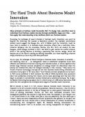
* $‘ he classical model of coagulation describes a ‘casQ cade’ of reactions involving activation of various clota ting factors along either an extrinsic or an intrinsic pathway. According to this model, stimulation of either of these two pathways can result in the production of a large amount of thrombin and subsequent formation of a fibrin clot’ (Fig. 1). However, although this cascade paradigm supports laboratory evaluation of coagulation disorders and demonstrates the interactions between coagulation factors, it does not adequately explain the mechanisms leading to haemostasis in viva. Furthermore, it does not provide a great deal of information regarding the pathophysiology of the haemostatic system. In particular, the model does not explain why certain categories of patients demonstrate a haemorrhagic tendency; nor does it facilitate accurare prediction of which patients will actually bleed. For instance, patients with a deficiency of factor XII @XII), high-molecular-weight kininogen, or prekallikrein do not present with a bleeding tendency de-
X
1
=
factor xa factor
: : : : _ i ; :
hypothesis:
intrinsic
4
facmr x
Va
fibrinogen Fig. I. The ‘cascade’
pathway
factor Vita Tissue facbr
Medical Center, Durham. North
Carolina. USA
INTRODUCTION
XIa
--+
and extrinsic
fibrin pathways.
: : : : :
spite a prolonged partial thromboplastin time (FIT), which indicates a disturbance in the functional activity of the intrinsic pathway.* In contrast, an increased predisposition to haemorrhagic risk may be present in patients deficient in FXI. The degree of prolongation of the FIT in this disorder, however, does not necessarily predict the extent of the bleeding tendency, which is typically less severe than that observed in haemophilia. The cascade hypothesis cannot account for the varying degrees of haemorrhagic tendency and diverse clinical observations that result from deficiencies of different components of the two pathways. In an attempt to explore the process of haemostasis from new angles, we developed experimental and conceptual models that would allow us to test hypotheses in a biochemical or Ed r&o system. This, in turn, would increase understanding of how the normal haemostatic system actually works in &JO. In addition, we wished to explore the mechanism of haemostasis in haemophiliac patients. Such patients have a normal prothrombin time (F’T), which measures activity of the extrinsic pathway, despite a prolonged F”IT and a pronounced bleeding tendency. Why, then, does the extrinsic pathway fail to compensate for the dysfunctional intrinsic pathway? In other words, why do haemophiliacs bleed? We have developed a cell-based model of haemostasis that will replace the classical model of the coagulation cascade.3 This cell-based model emphasises the interaction of clotting factors with specific cell surfaces4 and appears to be able to shed light on many of the unresolved issues highlighted by the traditional cascade theory.
;
THE
: : : : ! ! f : : : : : : I j
CELL-BASED
MODEL
OF
HAEMOSTASIS
The first step in our investigation was to establish an in vitro experimental system incorporating platelets and plasma concentrations of various clotting factors and coagulation inhibitors. A cellular source of tissue factor (TF) was considered to be essential, and inclusion of TF-bearing monocytes
0 2003 Elsevler Sdence Ltd. All rights reserved.
Blood Reviews
(2003) 17, 51-55
Table I
Cell-based
model
system Concentration
Component
(nM)
Cells Monocytes Platelets
-
(cultured
with agents
to induce
TF) -
(unactivated)
Proteins Prothrombin
I400
Factor
V
25
Factor
VIII
0.4
Factor
IX
70
Factor
X
135
Factor
XI
30
Factor
Vlla
0.2
’ IX Fig. 2. Haemostasis platelets.
Inhibitors
HR.
TFPI
3
ATIII
2500
TF, tissue factor;
TFPI, tissue
factor
pathway
inhibitor;
ATIII. antithrombin
occurs
(Reproduced
Activated
factor
activated
platelets:
activated
factor
565, with
permission.)
from
on two
VII activates thoughts
VII. Blood
cell surfaces:
Hoffman on the Coagul
TF-bearing
M., Monroe
factors
cells
3rd.
IX and X on the
mechanism Fibrinolysis
D.M.
of action 1998;
and
Roberts surface
of
of high-dose
9 (Suppl.
I): S6l-
Ill.
ensured that the system contained all necessary components of the haemostatic process (Table 1).5 The concepts and theories leading to the cell-based model of haemostasis all evolved from this kind of experimental system. We now know that haemostasis occurs on cell surfaces. Earlier theories derived from the cascade hypothesis suggested that the coagulation factors themselves were responsible for controlling haemostasis in a system where cells merely provided a phosphatidylserine-containing surface on which the procoagulant complexes could be assembled. However, the cell-based model proposes that cells play very active roles in controlling coagulation,* with certain features of the cell surfaces directing the haemostatic process. In this system, cells with similar phosphatidylserine contents are able to play very different roles depending on their complement of surface receptors.4 The cell-based model also emphasises that coagulation occurs in a series of three overlapping steps that take place on different cell surfaces, rather than as a cascade that produces an abundance of activated factors and inevitably leads to clot formation. The first phase, or initiation, occurs on a TF-bearing cell. In the amplification phase, platelets and cofactors are activated in order to prepare for large-scale thrombin generation. Finally, propagation occurs on the surface of platelets, and results in the production of large amounts of thrombin (Fig. 2).4 Initiation Coagulation is initiated on a TF-bearing cell, which produces the first activated factors (Fig. 3a). This TF pathway may still be referred to as an ‘extrinsic’ pathway, as the TF-bearing cell is, under normal circumstances, outside of the vascular system and therefore extrinsic to the blood. A large number of cells ekpress TP including stromal fibroblasts, mononuclear cells: macrophages and endothelial cells, but TF is not usually in contact with the blood until injury or inllammation occurs.
Blood Reviews
(2003) 17, 5 l-55
0 2003 Ekevier Science Ltd. All rights reserved.
However, reliable evidence exists suggesting that the reactions responsible for initiating coagulation occur all the time outside the vasculature in healthy individuals. Coagulation factors, including FVII, FX, and prothrombin are able to percolate through tissue spaces, and can leave the vasculature in amounts dependent on their molecular size. These factors can be detected in the lymph and assayed along with their activated forms and activation peptides. Based on this observation, an ‘idling’ theory has been proposed, in which the TF pathway remains constantly active, generating low levels of activated factors in the basal state.6 Therefore, continual production of small amounts of thrombin takes place outside the vasculature in healthy individuals, even under normal circumstances when vascular integrity remains intact. In effect, the initiation step of coagulation is proceeding at all times, but does not lead to formation of a blood clot as its location is separated from other key components of the coagulation system by an intact vessel wall. Amplification As a result of vessel damage, components of the haemostatic system that are normally unable to leave the vasculature due to their large size are now able to do so. The most important of these elements are platelets, PVIII, and von Wfflebrand factor (vWP). As they leave the vascular system, they come into contact with the limited amount of thrombin that is being generated on the surface of the TF-bearing cell. Platelets stick to the site of injury, forming a plug at the damaged vessel wall, and become fully activated by the thrombin. This same thrombin is also critically important in activating coagulation factors. It completes the activation of w, which is released from activated platelets, and is responsible for the cleavage and subsequent activation of FVIII frc%fn vWF (Fig. 3b). In addition, studies have shown that thrombin can also activate FXI, which binds to high-affinity sites on the surface of activated platelets.7,8 This may explain why FXII and other contact factors are not always necessary for
Ila
(4
IXa
+ FreevWF
TF
(b)
(--_______
TFPI
Fig. 3. The ceil-based
I I I I I A
model
of haemostasis:
coagulation, as initially postulated by the original cascade hypothesis. Although insufficient to result in clot formation by itself, the small amount of thrombin generated at the surface of TF-bearing cells during the initiation phase is essential in amplifying the procoagulant signal. At ;he end of the amplification phase, platelets activated by this limited amount of thrombin are clad in activated cofactors and FXIa, and the process of haemostasis moves into the propagation phase.
(a) initiation,
= tissue
factor
(b) amplification,
pathway
inhibitor.
(c) propagation.
Propagation During propagation, FlXa combines with its cofactor, FVIIIa, on the surface of activated platelets. Some of the required FIXa is produced on the surface of TF-bearing cells by TF/FVIIa, and can diffuse to the activated platelets as it is not inhibited by tissue factor pathway inhibitor (TPPI), and is only slowly inhibited by antithrombln III (ATIII). Factor IXa can also be produced on the platelet surface by FXIa. Once formed, the FIXa/FVIIIa complex activates FX to
0 2003 Elsevier Science ltd. All rights reserved.
Blood Reviews
(2003) I7, S/-S5
FXa, which immediately combines with its cofactor (Fig. 3~). The FXa/FVa complex then converts large amounts of prothrombin to thrombin, resulting in the cleavage of fibrinogen to fibrin monomers, which polymerise to consolidate the initial platelet plug into a stable fibrin clot. The cell-based model therefore places an emphasis on the cellular control of coagulation, and is subsequently able to explain some clinical aspects of haemostasis that the classical cascade hypothesis cannot.4 It allows a more thorough understanding of how the coagulation process works in viva, and provides a greater degree of consistency with clinical observations of various coagulation disorders,
WHY DO HAEMOPHILIACS
BLEED?
When compared to the traditional cascade theory, the cellbased model facilitates a greater understanding of the pathophysiological mechanisms leading to haemophilia. For instance, the cascade model does not explain why the extrinsic pathway appears unable to produce sufficient amounts of FX to at least partially compensate for a deficiency of FVIII or FIX. In other words, why does activation of FX by the TP/FVIIa complex fail to substitute for the FXa that would normally be generated by FIXa/PVIIIa?’ The cell-based model does not suggest that FXa generation by the TF/FVIIa complex is insufficient in haemophilia, but that it occurs on the wrong cell surface. The FIXa/FVIIIa complex activates FX on the surface of platelets during the propagation phase, whereas TF/PVIIa can only produce FXa on the surface of the TF-bearing cell. The FXa produced on the TF-bearing cell is unable to move to the activated platelet surface, as there exist two very efficient inhibitors of FXa in the plasma: TFPI and ATIII. At normal plasma levels, both TFPI and ATIII inhibit FXa so rapidly and effectively that the half-life of FXa is 1 minute or less in the fluid phase.2 Therefore, FXa that remains at the TF-bearing cell is relatively protected from inhibition, whereas any FXa that diffuses from the surface is rapidly inhibited. Accordingly, the cell-based model proposes that haemophilia is specifically a failure of platelet-surface FXa generation, which results in a lack of platelet-surface thrombin production.’ Haemophiliac patients demonstrate relatively normal initiation and amplification phases of coagulation, and so are able to form an initial platelet plug at the bleeding site, but they cannot generate the burst of thrombin at the platelet surface that is necessary to stabilise the initial plug into a fibrin clot.
HOW DOES HIGH-DOSE FVBa ENHANCE HAEMOSTASIS IN HAEMOPHJLIA? As discussed above, the cell-based model of coagulation suggests that the total amount of FXa produced is less important than the location in which it is generated.* We believe that FXa must be formed on the platelet surface by FIXa/PVIIIa, in close proximity to w, in order to be incorporated into prothrombinase complexes. This means that the TF/FVIIa complex cannot compensate for a lack of
m
Blood Reviews (2003) 17, S I-55
0 2003 Elsevier Science Ltd. All rights reserved.
FIX/FVIII, as it makes FXa in the wrong place. If this is the case, then efficient haemophilia treatment must involve the restoration of FXa generation on the platelet surface. Our data imply that high-dose FVIIa is able to do just that - it can enhance haemostasis in haemophiliacs by activating sufficient PX on the surface of activated platelets to support a burst of thrombin generation.’ OriginaIly, our group favoured a TFdependent mechanism in which high doses of FVIIa could ‘drive’ the TF pathway in haemophiliacs, enhancing the performance of the extrinsic pathway and therefore producing haemostasis. It is well recognised that PVIIa exhibits very little proteolytic activity in the absence of TE However, the doses of FVIIa required to achieve coagulation in haemophiliacs produced plasma levels that were several orders of magnitude greater than the & for binding of PVIIa to TF, leading some researchers to suggest that FVIIa is unlikely to work through a TFdependent mechanism.’ We used our experimental model to determine how high-dose FVIIa supports haemostasis in patients with haemophilia. It was found that FVIIa binds weakly to activated platelets, even though platelets do not carry TE Once bound to the platelet, PVIIa generates a small amount of PXa, leading to the production of a limited amount of thrombin on the platelet surface. These findings are also consistent with our conceptual model of coagulation, which postulates that platelet-surface FXa generation is required for the assembly of the prothrombinase complex and subsequent thrombin generation. Furthermore, the concentration of FVIIa required to produce detectable thrombin generation correlates with the lowest concentration of PVIIa necessary for clinical efficacy in haemophilia patients. lo When compared to the amount of FXa that would usually be produced by the FIXa/FVIIIa complex, the quantity generated by platelet-bound FVIIa is low. However, it is significantly higher than the level of FXa normally produced on platelets of haemophiliacs, and is certainly sufficient to enhance thrombin generation in experimental models of FIX and FVIII deficiency. We believe that haemophilia is characterised primarily by a faihrre of platelet-surface thrombin generation. If this is the case, then results from our studies in experimental models suggest that high levels of FVIIa may partially restore FXa generation on the platelet surface, leading to enhanced thrombin production in the absence of FIX or FVIII (Pig. 4). We have tentatively made two extrapolations of our in vitro data to the in vivo effects of high-dose FVIIa therapy in haemophilia. First, OUT data suggest that a high dose is needed because PVIIa binds to platelets with a low affinity o(d of 50-100 nM, rather than 5 1 nM or less for FVIIa binding to TF).2 As a result, a high concentration of FVIIa is required to achieve even a modest degree of platelet binding. At the concentrations of PVIIa attained in z&o, binding to platelets is not saturated. This observation led us to predict that an escalation of FVIIa dose should therefore increase platelet-surface thrombin generation. Several groUps have confirmed this theory by demonstrating that clinical efficacy may be attained by increasing the dose of FVIIa in those haemophikac patients who fail to respond to initial dose recommendations.
Fig. 4. High-dose eration
3rd, Roberts surface
FVlla partially
in haemophilia.
H.R. Activated
of activated
high-dose
activated
I): S6 IS65,
with
restores
(Reproduced factor
platelets: factor
Hoffman
VII activates
thoughts
VII. Blood
platelet-surface from
factors
on the Coagul
thrombin
gen-
M., Monroe
D.M.
IX and X on the
mechanism
Fibrinolysis
of action
of
1998; 9 (Suppl.
permission.)
The second extrapolation from studies of FVIIa in the experimental model is that the action of high-dose FVIIa in vivo is not directly dependent on n, but is instead plateletdependent. Earlier theories postulating a TFdependent mode of action for FVIIa explain the localisation of FVIIa a$ti$ity to the injury site, which may account for the relative lack of thrombotic complications observed during highdose FVIIa therapy,’ but do not adequately justify the requirement for high doses. lo However, a platelet-dependent mechanism in which FVIIa binds to platelets with low aflinity allows not only foe the localisation of FVIIa activity, but also explains why high doses are required to attain clinically effective levels of thrombin generation. While this theory of platelet dependence does not preclude other actions and effects of FVIIa, it is consistent with empirically determined dosing requirements. However, it is important to be aware that this mechanism is not truly TF-independent, as TF is still required for the initiation of coagulation. The theory of a plateletdependent mechanism of action simply implies that the primary effect of FVIIa occurs on the platelet surface. i
haemostatic process in vivo, and facilitates a greater understanding of the pathophysiological mechanisms behind coagulation disorders such as haemophilia. Haemophilia may be chamcterised by a failure of plateletsurface thrombin generation in the final propagation stage of the haemostatic process. High-dose recombinant FVIIa (rFVIIa; NovoSeven@, Novo Nordisk, Copenhagen, Denmark) has shown considerable success as a therapy for haemophiliacs and inhibitor patients, and this success may be due to a mechanism of action involving platelet-surface FXa generation. This results in enhanced thrombin production, and may partially compensate for the deficiency of FIX or FVIII. The efficacy of high-dose rFVIIa in haemophilia and Inhibitor patients has led to its use in a growing number of alternative indications, and data regarding its mechanism of action in such circumstances are scarce. However, we believe that even in the presence of the FIXa/FVIIIa complex, FVIIa may be able to enhance both FXa and FIXa levels on the platelet surface, thus augmenting the production of vital thrombin.
References I.
Davie EW, Ratnoff ting. Science
2.
Hoffman
Bloodline
Veldman
system
recombinant 4. 5.
Hoffman Monroe
DM.
88: 364-37 6.
When compared to the traditional cascade hypothesis, we believe that the cell-based conceptual model of haemostasis allows a more fundamental understanding of the clinical problems observed in some coagulation disorders by focusing on the central role of specific cell surfaces in controlling and directing the haemostatic process. Our cell-based model builds upon the foundations laid by the traditional cascade theory, but places greater emphasis on the roles of specific receptors present on the surfaces of the cells involved. Importantly, the cell-based model suggests that understanding the structure and function of coagulation proteins is necessary, but not sufficient, to understand haemostasis in vivo. Accordingly, this more recent model provides a potentially more accurate representation of the
Mann
KG.
Roberts
]A, Monroe of factor Thromb
high-dose
into the coagoptions
IO: 797-81
model
M. Platelet system.
for
the
KO. Li CQ,
with I.
of hemostasis.
procoagulant
com-
Br J Haematol
1994;
DM,
lb-IX-V
Roberts
An
HR.
Hoffman
19: 170-l
M. Oliver
I662-
factor
1668.
MR. Feedback of factor
acti-
XII. Arte-
77.
HR. Activated of activated
of high-dose
1998; 9 (Suppl
Xl bind-
promotes
2002; 277:
DM 3rd. Roberts of action
PN. Factor
in the absence
Vast Biol 1999;
DM, Hoffman
of thrombosis.
complex
J Biol Chem
XI on platelets
Fibrinolysis factor
diagnosis
Lopez JA, Walsh
IX and X on the surface
on the mechanism Monroe
2003;
A cell-based
glycoprotein
M, Monroe
vates factors
IO.
a cell-based
1992; 2: 365-370.
by thrombin.
Oliver
Coagul
analytes
Epidemiol
vation Hoffman
3rd.
insights
therapeutic
Med Chem
HR. Hoffman
Potential
Baglia FA, Badellino
rioscler
new
in a tissue factor-initiated
Ann
Xl activation
9.
clot-
2001; 85: 958-965.
ing to the platelet 8.
blood
I.
overview. 7.
DM
S. New for
Vlla. Curr
M, Monroe Haemost
intrinsic
I : 5-6.
M, Ehrenforth
factor
Thromb
for
of NovoSeven@using
2002;
and implications
plex assembly
CONCLUSIONS
of action
Reviews
A. Hoffman
ulation
sequence
145: I 3 IO- I 3 12.
M. Mechanism
model. 3.
OD. Waterfall
1964;
factor
VII acti-
platelets:
thoughts
factor
VII. Blood
activated
I): S6 I-S65. ]A, Roberts
Vlla is independent
of tissue
HR. Platelet factor.
activity
of
Br J Haematol
1997; 99: 542-547.
0 2003 Elsevier Science Ltd. All rights mserved.
Blood Reviews
(2003) 17, 5 l-55
m

Related documents

0 Pages • 3,516 Words • PDF • 1.5 MB

15 Pages • 7,327 Words • PDF • 183 KB

528 Pages • 134,748 Words • PDF • 2.4 MB

11 Pages • 10,287 Words • PDF • 224.8 KB

12 Pages • 7,162 Words • PDF • 354.1 KB

9 Pages • 6,323 Words • PDF • 205.7 KB
14 Pages • 6,133 Words • PDF • 580.8 KB

6 Pages • 3,340 Words • PDF • 504.2 KB

202 Pages • 64,190 Words • PDF • 1.7 MB

10 Pages • 223 Words • PDF • 449.8 KB

10 Pages • 5,288 Words • PDF • 64.4 KB

3 Pages • 520 Words • PDF • 489.6 KB