Biology of Microorganisms 13th ed. - M. Madigan, et al., (Pearson, 2012) BBS
This document was submitted by our user and they confirm that they have the consent to share it. Assuming that you are writer or own the copyright of this document, report to us by using this DMCA report button.
Archaeoglobus
Halococcus Extreme halophiles
Marine Crenarchaeota
Euryarchaeota
Natronococcus Methanobacterium
Methanocaldococcus
Crenarchaeota
Halophilic methanogen
Methanothermus Sulfolobus Pyrodictium
Methanosarcina
Thermococcus/ Pyrococcus
Methanospirillum
Nanoarchaeum
Thermoplasma
Thermoproteus
Methanopyrus Desulfurococcus Picrophilus
Ferroplasma Root Extreme acidophiles
Hyperthermophiles
Protists Stramenopiles Oomycetes Brown Diatoms algae Golden Radioalgae Ciliates Alveolates Cercozoans Chlorarach- larians niophytes Dinoflagellates ForaminApicomplexans iferans Parabasalids Red algae Diplomonads (Secondary Green endosymbioses) Kinetoplastids algae Plants
Euglenids
Euglenozoa
Cellular slime molds Plasmodial slime molds Entamoebas Amoebozoa Gymnamoebas Chloroplast ancestor (primary endosymbiosis)
Bacteria
Animals Mitochondrial ancestor (primary endosymbiosis)
Fungi Microsporidia
Fungi
Brock
Biology of Microorganisms Thirteenth Edition
Michael T. Madigan Southern Illinois University Carbondale
John M. Martinko Southern Illinois University Carbondale
David A. Stahl University of Washington Seattle
David P. Clark Southern Illinois University Carbondale
Executive Editor: Deirdre Espinoza Project Editor: Katie Cook Associate Project Editor: Shannon Cutt Development Editor: Elmarie Hutchinson Art Development Manager: Laura Southworth Art Editor: Elisheva Marcus Managing Editor: Deborah Cogan Production Manager: Michele Mangelli Production Supervisor: Karen Gulliver Copyeditor: Anita Wagner
Art Coordinator: Jean Lake Photo Researcher: Maureen Spuhler Director, Media Development: Lauren Fogel Media Producers: Sarah Young-Dualan, Lucinda Bingham, and Ziki Dekel Art: Imagineering Media Services, Inc. Text Design: Riezebos Holzbaur Design Group Senior Manufacturing Buyer: Stacey Weinberger Senior Marketing Manager: Neena Bali Compositor: Progressive Information Technologies Cover Design: Riezebos Holzbaur Design Group
Cover Image: (front cover) Peter Siver/Visuals Unlimited/Corbis; (back cover) J.-H. Becking, Wageningen Agricultural University, Wageningen, Netherlands Credits for selected images can be found on page P-1.
Copyright © 2012, 2009, 2006 Pearson Education, Inc., publishing as Benjamin Cummings, 1301 Sansome Street, San Francisco, CA 94111. All rights reserved. Manufactured in the United States of America. This publication is protected by copyright and permission should be obtained from the publisher prior to any prohibited reproduction, storage in a retrieval system, or transmission in any form or by any means, electronic, mechanical, photocopying, recording, or likewise. To obtain permission(s) to use material from this work, please submit a written request to Pearson Education, Inc., Permissions Department, 1900 E. Lake Ave., Glenview, IL 60025. For information regarding permissions, call (847) 486-2635. Many of the designations used by manufacturers and sellers to distinguish their products are claimed as trademarks. Where those designations appear in this book, and the publisher was aware of a trademark claim, the designations have been printed in initial caps or all caps. Library of Congress Cataloging-in-Publication Data Brock biology of microorganisms / Michael T. Madigan . . . [et al.].—13th ed. p. cm. Includes index. ISBN-13: 978-0-321-64963-8 (hardcover : alk. paper) ISBN-10: 0-321-64963-X (hardcover : alk. paper) 1. Microbiology. I. Madigan, Michael T., 1949– II. Title: Biology of microorganisms. QR41.2.B77 2011 579—dc22 2010044962
ISBN 10: 0-321-64963-X (student edition) ISBN 13: 978-0-321-64963-8 (student edition) ISBN 10: 0-321-72675-8 (professional copy) ISBN 13: 978-0-321-72675-9 (professional copy) 1 2 3 4 5 6 7 8 9 10—CRK—14 13 12 11 10
About the Authors
Michael T. Madigan
received his B.S. in Biology and Education from Wisconsin State University–Stevens Point (1971) and his M.S. (1974) and Ph.D. (1976) in Bacteriology from the University of Wisconsin–Madison. His graduate research was on the hot spring bacterium Chloroflexus in the laboratory of Thomas Brock. Following a three-year postdoctoral in the Department of Microbiology, Indiana University, Mike moved to Southern Illinois University– Carbondale, where he has been a professor of microbiology for 32 years. He has coauthored Biology of Microorganisms since the fourth edition (1984) and teaches courses in introductory microbiology, bacterial diversity, and diagnostic and applied microbiology. In 1988 Mike was selected as the Outstanding Teacher in the College of Science and in 1993, the Outstanding Researcher. In 2001 he received the SIUC Outstanding Scholar Award. In 2003 he received the Carski Award for Distinguished Undergraduate Teaching from the American Society for Microbiology (ASM), and he is an elected Fellow of the American Academy of Microbiology. Mike’s research is focused on bacteria that inhabit extreme environments, and for the past 12 years he has studied the microbiology of permanently ice-covered lakes in the McMurdo Dry Valleys, Antarctica. In addition to his research papers, he has edited a major treatise on phototrophic bacteria and served for over a decade as chief editor of the journal Archives of Microbiology. He currently serves on the editorial board of the journals Environmental Microbiology and Antonie van Leeuwenhoek. Mike’s nonscientific interests include forestry, reading, and caring for his dogs and horses. He lives beside a peaceful and quiet lake with his wife, Nancy, five shelter dogs (Gaino, Snuffy, Pepto, Peanut, and Merry), and four horses (Springer, Feivel, Gwen, and Festus).
John M. Martinko received his B.S. in Biology from Cleveland State University. He then worked at Case Western Reserve University, conducting research on the serology and epidemiology of Streptococcus pyogenes. His doctoral work at the State University of New York–Buffalo investigated antibody specificity and antibody idiotypes. As a postdoctoral fellow, he worked at Albert Einstein College of Medicine in New York on the structure of major histocompatibility complex proteins. Since 1981, he has been in the Department of Microbiology at Southern Illinois University–Carbondale where he was Associate Professor and Chair, and Director of the Molecular Biology, Microbiology, and Biochemistry Graduate Program. He retired in 2009, but remains active in the department as a researcher and teacher. His research investigates structural changes in major histocompatibility proteins. He teaches an advanced course in immunology and presents immunology and host defense lectures to medical students. He also chairs the Institutional Animal Care and Use Committee at SIUC. He has been active in educational outreach programs for pre-university students and teachers. For his educational efforts, he won the 2007 SIUC Outstanding Teaching Award. He is also an avid golfer and cyclist. John lives in Carbondale with his wife Judy, a high school science teacher.
iii
iv
About the Authors
David A. Stahl received his B.S. degree in Microbiology from the University of Washington–Seattle, later completing graduate studies in microbial phylogeny and evolution with Carl Woese in the Department of Microbiology at the University of Illinois–Champaign-Urbana. Subsequent work as a postdoctoral fellow with Norman Pace, then at the National Jewish Hospital in Colorado, focused on early applications of 16S rRNA-based sequence analysis to the study of natural microbial communities. In 1984 Dave joined the faculty at the University of Illinois–Champaign-Urbana, holding appointments in Veterinary Medicine, Microbiology, and Civil Engineering. In 1994 he moved to the Department of Civil Engineering at Northwestern University, and in 2000 returned to his alma mater, the University of Washington–Seattle, as a professor in the Departments of Civil and Environmental Engineering and Microbiology. Dave is known for his work in microbial evolution, ecology, and systematics, and received the 1999 Bergey Award and the 2006 Procter & Gamble Award in Applied and Environmental Microbiology from the ASM; he is also an elected Fellow of the American Academy of Microbiology. His main research interests are the biogeochemistry of nitrogen and sulfur compounds and the microbial communities that sustain these nutrient cycles. His laboratory was first to culture ammonia-oxidizing Archaea, a group now believed to be the main mediators of this key process in the nitrogen cycle. He has taught several courses in environmental microbiology, is one of the co-founding editors of the journal Environmental Microbiology, and has served on many advisory committees. Outside teaching and the lab, Dave enjoys hiking, bicycling, spending time with family, reading a good science fiction book, and, with his wife Lin, renovating an old farmhouse on Bainbridge Island, Washington.
Dedications Michael T. Madigan dedicates this book to the memory of his children who rest on Boot Hill: Andy, Marcy, Willie, Plum, Teal, and Sugar. Whether in good times or bad, they always greeted him with tails a waggin’.
John M. Martinko dedicates this book to his daughters Sarah, Helen, and Martha, and to his wife Judy. Thanks for all of your support!
David A. Stahl dedicates this book to his wife, Lin. My love, and one that helps me keep the important things in perspective.
David P. Clark dedicates this book to his father, Leslie, who set him the example of reading as many books as possible.
David P. Clark
grew up in Croydon, a London suburb. He won a scholarship to Christ’s College, Cambridge, where he received his B.A. degree in Natural Sciences in 1973. In 1977 he received his Ph.D. from Bristol University, Department of Bacteriology, for work on the effect of cell envelope composition on the entry of antibiotics into Escherichia coli. He then left England on a postdoctoral studying the genetics of lipid metabolism in the laboratory of John Cronan at Yale University. A year later he moved with the same laboratory to the University of Illinois at Urbana-Champaign. David joined the Department of Microbiology at Southern Illinois University–Carbondale in 1981. His research has focused on the growth of bacteria by fermentation under anaerobic conditions. He has published numerous research papers and graduated over 20 Masters and Doctoral students. In 1989 he won the SIUC College of Science Outstanding Researcher Award. In 1991 he was the Royal Society Guest Research Fellow at the Department of Molecular Biology and Biotechnology, Sheffield University, England. In addition to Brock Biology of Microorganisms, David is the author of four other science books: Molecular Biology Made Simple and Fun, now in its fourth edition; Molecular Biology: Understanding the Genetic Revolution; Biotechnology: Applying the Genetic Revolution; and Germs, Genes, & Civilization: How Epidemics Shaped Who We Are Today. David is unmarried and lives with two cats, Little George, who is orange and very nosey, and Mr. Ralph, who is mostly black and eats cardboard.
Preface
T
he authors and Benjamin Cummings Publishers proudly present the 13th edition of Brock Biology of Microorganisms (BBOM 13/e). This book is truly a milestone in the annals of microbiology textbooks. Brock Biology of Microorganisms, and its predecessor, Biology of Microorganisms, has introduced the field of microbiology to students for 41 years, more than any other textbook of microbiology. Nevertheless, although this book goes back over four decades, its two main objectives have remained firm since the first edition was published in 1970: (1) to present the principles of microbiology in a clear and engaging fashion, and (2) to provide the classroom tools necessary for delivering outstanding microbiology courses. The 13th edition of BBOM fulfills these objectives in new and exciting ways. Veteran textbook authors Madigan, Martinko, and Clark welcome our new coauthor, Dave Stahl, to this edition of BBOM. Dave is one of the world’s foremost experts in microbial ecology and has masterfully crafted an exciting new view of the ecology material in BBOM, including a new chapter devoted entirely to microbial symbioses, a first for any textbook of microbiology. Users will find that the themes of ecology and evolution that have permeated this book since its inception reach new heights in the 13th edition. These fundamental themes also underlie the remaining content of the book—the basic principles of microbiology, the molecular biology and genetics that support microbiology today, the huge diversity of metabolisms and organisms, and the medical and immunological facets of microbiology. It is our belief that outstanding content coupled with outstanding presentation have come together to make BBOM 13/e the most comprehensive and effective textbook of microbiology available today.
key concepts from each numbered section in a wrap-up style that is certain to be a big hit with students, especially the night before examinations! Our end-of-chapter key terms list, two detailed appendices, a comprehensive glossary, and a thorough index complete the hard copy learning package. Many additional learning resources are available online (see below). In terms of presentation, BBOM 13/e will easily draw in and engage the reader. The book has been designed in a beautiful yet simple fashion that gives the art and pedagogical elements the breathing room they need to be effective and the authors the freedom to present concepts in a more visually appealing way. Supporting the narrative are spectacular illustrations, with every piece of art rendered in a refreshing new style. Moreover, the art complements, and in many cases integrates, the hundreds of photos in BBOM, many of which are new to the 13th edition. And, as users of BBOM have come to expect, our distinctive illustrations remain the most accurate and consistent of those in any microbiology textbook today. The authors are keenly aware that it is easy to keep piling on new material and fattening up a textbook. In response to this trend, BBOM 13/e went on a diet. With careful attention to content and presentation, BBOM 13/e is actually a shorter book than BBOM 12/e. The authors have carefully considered every topic to ensure that content at any point in the book is a reflection of both what the student already knows and what the student needs to know in a world where microbiology has become the most exciting and relevant of the biological sciences. The result is a more streamlined and exciting treatment of microbiology that both students and instructors will appreciate.
What’s New in the 13th Edition?
Revision Highlights:
In terms of content and pedagogy, instructors who have used BBOM previously will find the 13th edition to be the same old friend they remember; that is, a book loaded with accurate, upto-the-minute content that is impeccably organized and visually enticing. The 36 chapters in BBOM 13/e are organized into modules by numbered head, which allows instructors to fine-tune course content to the needs of their students. In addition, study aids and review tools are an integral part of the text. Our new MiniQuiz feature, which debuts in the 13th edition, is designed to quiz students’ comprehension as they work their way through each chapter. Also new to this edition is the end-of-chapter review tool called “Big Ideas.” These capsule summaries pull together the
Chapter 1 • Find new coverage on the evolution and major habitats of microorganisms—Earth’s most pervasive and extensive biomass. • A more visually compelling presentation of the impacts of microorganisms on humans better emphasizes the importance of microorganisms for the maintenance of all life on Earth.
Chapter 2 • New coverage of cell biology and the nature of the chromosome in prokaryotic and eukaryotic cells is complemented by a visually engaging overview of the microbial world. v
vi
Preface
Chapter 3
Chapter 10
• The cell chemistry chapter that previously held this position is now available online (www.microbiologyplace.com). The new Chapter 3 explores cell structure and function with strong new visuals to carry the text and new coverage of the lipids and cell walls of Bacteria and Archaea.
• The fundamental principles of microbial genetics are updated and supplemented with new coverage that compares and contrasts bacterial and archaeal genetics.
Chapter 4
• Find “one-stop shopping” for coverage of molecular biological methods, including cloning and genetic manipulations, as a prelude to the genomics discussion in the next chapter. • Enjoy the colorful new Microbial Sidebar on new fluorescent labeling methods that can differentiate even very closely related bacteria.
• Find updated coverage of catabolic principles along with an overview of essential anabolic reactions. • Newly rendered and more instructive art makes mastering key metabolic pathways and bioenergetic principles a more visual experience.
Chapter 5 • Updated coverage of the events in cell division and their relation to medical microbiology connects basic science to applications. • Newly rendered art throughout makes the important concepts of cell division and population growth more vivid, engaging, and interactive.
Chapter 6 • The concise primer on molecular biology that every student needs to know is updated and now includes an overview of the structures of nucleic acids and proteins and the nature of chromosomes and plasmids.
Chapter 7 • Find new coverage of the latest discoveries in the molecular biology of Archaea and comparisons with related molecular processes in Bacteria. • A new section highlights the emerging area of regulation by microRNA in eukaryotes.
Chapter 8 • Review major updates on the regulation of gene expression— one of the hottest areas in microbiology today—including expanded coverage of cell sensing capacities and signal transduction. • Enjoy the new Microbial Sidebar featuring CRISPR, the newly discovered form of RNA-based regulation used by Bacteria and Archaea to ward off viral attack.
Chapter 9 • Major updates of the principles of virology are complemented with an overview of viral diversity. • New art reinforces the relevance and importance of viruses as agents of genetic exchange.
Chapter 11
Chapter 12 • Extensive updates on microbial genomics and transcriptomics will be found along with new coverage of the emerging related areas of metabolomics and interactomics. • Readers will marvel at the diversity of prokaryotic genomes in the new Microbial Sidebar “Record-Holding Bacterial Genomes.”
Chapter 13 • The two chapters covering metabolic diversity have been revised and moved up to Chapters 13 and 14 to precede rather than follow coverage of microbial diversity, better linking these two important and often related areas. • This chapter is loaded with reworked art and text that highlight the unity and diversity of the bioenergetics underlying phototrophic and chemolithotrophic metabolisms.
Chapter 14 • Restyled and impeccably consistent art showcases the comparative biochemistry of the aerobic and anaerobic catabolism of carbon compounds.
Chapter 15 • This retooled chapter combines the essentials of industrial microbiology and biotechnology, including the production of biofuels and emerging green microbial technologies.
Chapter 16 • Find new coverage of the origin of life and how the evolutionary process works in microorganisms. • Microbial phylogenies from small subunit ribosomal RNA gene analyses are compared with those from multiple-gene and full genomic analyses.
Preface
vii
Chapters 17–19
Chapter 25
• Coverage of the diversity of Bacteria and Archaea better emphasizes phylogeny with increased focus on phyla of particular importance to plants and animals and to the health of our planet. • Spectacular photomicrographs and electron micrographs carry the reader through prokaryotic diversity.
• This new chapter focuses entirely on microbial symbioses, including bacterial–bacterial symbioses and symbioses between bacteria and their plant, mammal, or invertebrate hosts. Find coverage here of all of the established as well as more recently discovered symbioses, including the human gut and how its microbiome may control obesity, the rumen of animals important to agriculture, the hindgut of termites, the light organ of the squid, the symbioses between hydrothermal vent animals and chemolithotrophic bacteria, the essential bacterial symbioses of insects, medicinal leeches, reef-building corals, and more, all supported by spectacular new color photos and art. • Learn how insects have shaped the genomes of their bacterial endosymbionts. • Marvel at the new Microbial Sidebar that tells the intriguing story of the attine ants and their fungal gardens.
Chapter 20 • A heavily revised treatment of the diversity of microbial eukaryotes is supported by many stunning new color photos and photomicrographs. • Find an increased emphasis on the phylogenetic relationships of eukaryotes and the “bacterial nature” of eukaryotic organelles.
Chapter 21 • Viruses, the most genetically diverse of all microorganisms, come into sharper focus with major updates on their diversity. • A new section describes viruses in nature and their abundance in aquatic habitats.
Chapter 22 • This chapter features a major new treatment of the latest molecular techniques used in microbial ecology, including CARD-FISH, ARISA, biosensors, NanoSIMS, flow cytometry, and multiple displacement DNA amplification. • Find exciting new coverage of methods for functional analyses of single cells, including single-cell genomics and single-cell stable isotope analysis, and expanded coverage of methods for analyses of microbial communities, including metagenomics, metatranscriptomics, and metaproteomics.
Chapter 23 • A comparison of the major habitats of Bacteria and Archaea is supported by spectacular new photos and by art that summarizes the phylogenetic diversity and functional significance of prokaryotes in each habitat. • Find broad new coverage of the microbial ecology of microbial mat communities and prokaryotes that inhabit the deep subsurface.
Chapter 24 • Revised coverage of the classical nutrient cycles is bolstered by new art, while new coverage highlights the calcium and silica cycles and how these affect CO2 sequestration and global climate. • Improved integration of biodegradation and bioremediation shows how natural microbial processes can be exploited for the benefit of humankind.
Chapter 26 • Key updates will be found on microbial drug resistance and are supported by new art that reveals the frightening reality that several human pathogens are resistant to all known antimicrobial drugs.
Chapter 27 • Extensively reworked sections on the normal microbial flora of humans include new coverage of the human microbiome and a molecular snapshot of the skin microflora. • Find revised coverage of the principles of virulence and pathogenicity that connect infection and disease.
Chapter 28 • Here we present the perfect overview of immunology for instructors who wish to cover only the fundamental concepts and how the immune system resists the onslaught of infectious disease. • Find late-breaking practical information on the immune response, including vaccines and immune allergies.
Chapter 29 • Built on the shoulders of the previous chapter, here is a more detailed probe of the mechanisms of immunity with emphasis on the molecular and cellular interactions that control innate and adaptive immunity.
Chapter 30 • This short chapter presents an exclusively molecular picture of immunology, including receptor–ligand interactions (the “triggers” of the immune response), along with genetics of the key proteins that drive adaptive immunity.
viii
Preface
Chapter 31
Chapter 34
• Find revised and expanded coverage of molecular analyses in clinical microbiology, including new enzyme immunoassays, reverse transcriptase PCR, and real-time PCR.
• Follow the emergence, rapid dispersal, and eventual entrenchment of West Nile virus as an endemic disease in North America. • Expanded coverage of malaria—the deadliest human disease of all time—includes the promise of new antiparasitic drugs and disease prevention methods.
Chapter 32 • Review major updates of the principles of disease tracking, using 2009 pandemic H1N1 influenza as a model for how newly emerging infectious diseases are tracked. • Find updated coverage throughout, especially of the HIV/AIDS pandemic.
Chapter 33 • Read all about the origins and history of pandemic H1N1 influenza and how the H1N1 virus is related to strains of influenza that already existed in animal populations. • Hot new coverage of immunization strategies for HIV/AIDS.
Chapter 35 • Find updates of water microbiology, including new rapid methods for detecting specific indicator organisms.
Chapter 36 • Explore new methods of food processing, including aseptic and high-pressure methods that can dramatically extend the shelflife and safety of perishable foods and drinks.
Cutting Edge Coverage Includes the Most Current Presentation of Microbial Ecology The 13th edition enhances the themes of ecology and evolution throughout, and is the only book on the market to include specialized coverage of archael and eukaryotic molecular biology. The book represents the most current research in the field, with special attention paid to the microbial ecology chapters:
Chapter 22, Methods in Microbial Ecology, is heavily updated to present the latest molecular techniques used in microbial ecology, including CARD-FISH, ARISA, biosensors, NanoSIMS, flow cytometry, and multiple displacement DNA amplification. It also includes exciting new coverage of methods for functional analyses of single cells, including single-cell genomics and single-cell stable isotope analysis, and expanded coverage of methods for analyses of microbial communities, including metagenomics, metatranscriptomics, and metaproteomics. Firmicutes Planctomycetes Cyanobacteria
Bacteroidetes Other
Chapter 23, Major Microbial Habitats and Diversity, compares the major habitats of Bacteria and Archaea and is supported by spectacular new photos and art that summarize the phylogenetic diversity and functional significance of prokaryotes in each habitat.
Burkholderiales Nitrosomonadales
`
Euryarchaeota Crenarchaeota
Rhodobacterales
_
Unclassified and minor bacterial groups
SAR11 group
a Other
Archaea
Proteobacteria
Alteromonadales
Oceanospirillales Pseudomonadales Vibrionales
Actinobacteria Acidobacteria Other Archaea
Other Proteobacteria
b
Verrucomicrobia
¡
Figure 23.24
Ocean prokaryotic diversity. The results are pooled analyses of 25,975 sequences from several studies of the 16S rRNA gene content of pelagic ocean waters. Many of these groups are covered in Chapters 17 and 18 (Bacteria) or 19 (Archaea). For Proteobacteria, major subgroups are indicated. Note the high proportion of cyanobacterial and Gammaproteobacteria sequences. Data assembled and analyzed by Nicolas Pinel.
Chapter 24, Nutrient Cycles, Biodegradation, and Bioremediation. Exciting updates of all the nutrient cycles that form the heart of environmental microbiology and
Bacteroidetes
microbial ecology.
Chapter 25, Microbial Symbioses, is a completely new chapter focused entirely on microbial symbioses,
Ochrobactrum
Yoshitomo Kikuchi and Jörg Graf
Betaproteobacteria
Figure 25.40 Micrograph of a FISH-stained microbial community in the bladder of Hirudo verbana. A probe (red) targeted at the 16S rRNA of Betaproteobacteria and a probe (green) targeted at the 16S rRNA of Bacteroidetes reveal distinct layers of different bacteria in the lumen of the bladder. Staining with DAPI (blue), which binds to DNA, reveals the intracellular alphaproteobacterium Ochrobactrum and host nuclei.
including bacterial–bacterial symbioses and symbioses between bacteria and their plant, mammal, or invertebrate hosts. Find coverage here of all the established as well as more recently discovered symbioses—including the human gut and how its microbiome may control obesity, the rumen of animals important to agriculture, the hindgut of termites, the light organ of the squid, the symbioses between hydrothermal vent animals and chemolithotrophic bacteria, and the essential bacterial symbioses of insects, medicinal leeches, reef-building corals, and more.
For a detailed list of chapter-by-chapter updates, see page v of the Preface. ix
Thoroughly Updated and Revised Art The art has been revised and updated throughout the book to give students a clear view into the microbial world. Color and style conventions are used consistently to make the art accessible and easy to understand.
Carefully redesigned new art clearly guides students through challenging concepts. The style for metabolic figures and other pathway processes has been simplified, and color-coded steps and chemical structures increase student comprehension.
Dimensionality has been added to some figures, lending more realism and vivacity to the presentation. Figures in which nucleic acids or cells are depicted are now more dimensional to clearly identify key genes and cell structures.
x
Illustrations and photos are often paired to give an idealized view next to a realistic view and to reinforce the connection between theory and practice.
xi
Conceptual Framework Helps Students Focus on the Key Concepts
The first twelve chapters cover the principles of microbiology. Basic principles are presented early on and then used as the foundation to tackle the material in greater detail later.
Brief Contents UNIT I
Basic Principles of Microbiology Chapter 1 Chapter 2 Chapter 3
Microorganisms and Microbiology A Brief Journey to the Microbial World Cell Structure and Function in Bacteria and Archaea
1 24 47
UNIT 2
Metabolism and Growth Chapter 4 Chapter 5
Nutrition, Culture, and Metabolism of Microorganisms Microbial Growth
85 117
Chapter 20 Eukaryotic Cell Biology and Eukaryotic Microorganisms Chapter 21 Viral Diversity
584 613
UNIT 7
Microbial Ecology Chapter 22 Methods in Microbial Ecology Chapter 23 Major Microbial Habitats and Diversity Chapter 24 Nutrient Cycles, Biodegradation, and Bioremediation Chapter 25 Microbial Symbioses
642 669 698 720
UNIT 8 UNIT 3
Molecular Biology and Gene Expression Chapter 6 Chapter 7 Chapter 8
Molecular Biology of Bacteria Archaeal and Eukaryotic Molecular Biology Regulation of Gene Expression
150 191 209
UNIT 4
Virology, Genetics, and Genomics Chapter 9 Chapter 10 Chapter 11 Chapter 12
Viruses and Virology Genetics of Bacteria and Archaea Genetic Engineering Microbial Genomics
236 263 291 313
UNIT 5
Microbial Evolution and Diversity Microbial Evolution and Systematics Bacteria: The Proteobacteria Other Bacteria Archaea
xviii
Information on metabolic diversity precedes the coverage of microbial diversity, better linking these important and often related areas.
xii
755 787
UNIT 9
Immunology Chapter 28 Immunity and Host Defense Chapter 29 Immune Mechanisms Chapter 30 Molecular Immunology
816 838 859
UNIT 10
Diagnosing and Tracking Microbial Diseases 878 913
UNIT 11 340 372 411
UNIT 6
Chapter 16 Chapter 17 Chapter 18 Chapter 19
Chapter 26 Microbial Growth Control Chapter 27 Microbial Interactions with Humans
Chapter 31 Diagnostic Microbiology and Immunology Chapter 32 Epidemiology
Metabolic Diversity and Commercial Biocatalyses Chapter 13 Phototrophy, Chemolithotrophy, and Major Biosyntheses Chapter 14 Catabolism of Organic Compounds Chapter 15 Commercial Products and Biotechnology
Antimicrobial Agents and Pathogenicity
446 475 517 556
New chapter on symbiosis ties together the core concepts of the book—health, diversity, and the human ecosystem.
Human- and Animal-Transmitted Infectious Diseases Chapter 33 Person-to-Person Microbial Diseases Chapter 34 Vectorborne and Soilborne Microbial Pathogens UNIT 12
944 981
Common-Source Infectious Disease Chapter 35 Wastewater Treatment, Water Purification, and Waterborne Microbial Diseases Chapter 36 Food Preservation and Foodborne Microbial Diseases
1004 1022
This newly revised chapter is the perfect overview for instructors who wish to cover immunology at a generalized level including the fundamental concepts of how the immune system resists the onslaught of infectious disease. Instructors who like to go into more detail can build on the core principles taught in Chapter 28 by covering Immune Mechanisms (Ch. 29) and Molecular Immunology (Ch. 30).
The new Big Ideas sections at the end of each chapter focus on the core concepts students need to know.
MiniQuiz MiniQuiz critical thinking questions integrated throughout the text test student comprehension of core principles from each section.
• What are the primary response regulator and the primary sensor kinase for regulating chemotaxis? • Why is adaptation during chemotaxis important? • How does the response of the chemotaxis system to an attractant differ from its response to a repellent?
xiii
Additional Resources
FOR STUDENTS
The MyMicrobiologyPlace website is rich with media assets to give students extra practice. It includes chapter quizzes, new quantitative questions, animations, and additional tutorials. www.microbiologyplace.com
Quantitative Questions
1
Number of genes in plasmid R100.
The Escherichia coli plasmid R100 is a circular molecule of DNA containing 93.4 kbp. The average E. coli protein contains 300 amino acids; assume that the same is true for R100 proteins. With this assumption, calculate how many genes are in this plasmid.
2 Compare DNA polymerases. Escherichia coli
FOR INSTRUCTORS
contains at least five different DNA polymerases. The three most characterized are DNA Pol I, Pol II, and Pol III. Polymerase I and II replicate DNA at about 20–40 nucleotides/sec whereas Pol III replicates at 250 to 1000 nucleotides/sec. The genome of E. coli strain K-12 is 4,639,221 bp. At the higher rates, how long does it take to reproduce the chromosome? How do these numbers agree with the roles of these DNA polymerases?
CourseCompass includes all of the assets from the MyMicrobiologyPlace website and all of the test questions from the computerized test bank. It also features class management tools, such as discussion boards and email functionality to help instructors easily teach online classes or give assignments. www.aw-bc.com/coursecompass
xiv
Instructor Resource DVD (IR-DVD)
Instructor Manual and Test Bank
0-321-72086-5 / 978-0-321-72086-3
0-321-72021-0 / 978-0-321-72021-4
The IR-DVD offers a wealth of media resources including all the art from the book in both JPEG and PPT formats, PowerPoint lecture outlines, computerized test bank, and answer keys all in one convenient location. The animations help bring lectures to life, while the select step-edit figures help break down complicated processes.
by W. Matthew Sattley and Christopher A. Gulvik The Instructor Manual/Test Bank provides chapter summaries that help with class preparation as well as the answers to the end-of-chapter review and application questions. The test bank contains 3,000 questions for use in quizzes, tests, and exams.
Acknowledgments book of this stature is not the product of its authors alone but instead is the collective effort of the many people who comprise the book team. These include folks both inside and outside of Benjamin Cummings. Executive editor Deirdre Espinoza and project editor Katie Cook, both of Benjamin Cummings, were the workhorses in editorial. Deirdre paved the way for the 13th edition and skillfully maneuvered the book around the occasional roadblocks that accompany any major textbook project. Katie ran the day-to-day operations of the BBOM team in a highly professional manner, expertly managing reviews and many other details and keeping all facets of the project on track. The BBOM 13/e production and design team was headed up by Michele Mangelli (Mangelli Productions) who oversaw Yvo Riezebos (Riezebos Holzbaur Design Group), and Laura Southworth (Benjamin Cummings). Michele managed the production team and did a great job of keeping everyone on mission and on budget. The artistic magic of Yvo is clearly visible in the beautiful text and cover designs of BBOM 13/e. Laura created the new art look for BBOM 13/e, one that readers should immediately appreciate for its clarity, consistency, and modern style. The authors are extremely grateful to Michele, Yvo, and Laura, as well as to the artist team at the studio of Imagineering (Toronto), for helping the authors produce such a beautiful book. Others in production included Karen Gulliver, Jean Lake, and Maureen Spuhler. Karen was our excellent production editor who ensured that a polished book emerged from a raw manuscript, while Jean was our art coordinator, tracking and routing art and handling interactions with the art studio. Maureen was our photo researcher who helped the authors locate photos that met the exacting standards of BBOM. The authors are extremely grateful to Karen, Jean, and Maureen for transforming literally thousands of pages of text and art manuscript into a superb learning tool. The authors wish to give special thanks to four other members of the production team: Elmarie Hutchinson, Anita Wagner, Elisheva (Ellie) Marcus, and Elizabeth McPherson. Our developmental editor Elmarie was a key contributor early in the project, helping the authors better link text and art and massaging the text to improve readability. Anita was our absolutely spectacular copyeditor; the authors could not have asked for a brighter or more effective person in this key position on the book team. Anita improved the accuracy, clarity, and consistency of the text and rendered her editorial services in a style that the authors found both helpful and time saving. Ellie (Benjamin Cummings) was our art liaison on this project, translating for the art house the intentions of the authors. Ellie has the unique gift of viewing art from both an artistic and a scientific perspective. Therefore, the consistency, clarity, and accuracy of the art in BBOM 13/e are
A
in large part due to her superb efforts. Elizabeth (University of Tennessee) was our manuscript accuracy checker; her eagle eye, extensive knowledge of all areas of microbiology, prompt service, and knack for editorial troubleshooting greatly improved the accuracy and authority of the final product. The authors also wish to acknowledge the excellent contributions of Dr. Matt Sattley, Indiana Wesleyan University. Matt, a former doctoral student of MTM, composed the Instructor’s Manual that accompanies BBOM 13/e. The manual should greatly assist instructors of any vintage to better organize their microbiology courses and select review questions for student assignments. We also thank Christopher Gulvik, University of Tennessee, for revising the test bank questions for this edition. No textbook in microbiology could be published without thorough reviewing of the manuscript and the gift of new photos from experts in the field. We are therefore extremely grateful for the kind help of the many individuals who provided general or technical reviews of the manuscript or who supplied new photos. They are listed below. And last but not least, the authors thank the women in their lives—Nancy (MTM), Judy (JMM), Linda (DAS), and Donna (DPC)—for the sacrifices they have made the past two years while this book was in preparation and for simply putting up with them during the ordeal that is “a BBOM revision.” F.C. Thomas Allnutt Daniel Arp, Oregon State University Marie Asao, Ohio State University Tracey Baas, University of Rochester Zsuzsanna Balogh-Brunstad, Hartwick College Teri Balser, University of Wisconsin–Madison Tamar Barkay, Rutgers University John Baross, University of Washington Douglas Bartlett, Scripps Institute of Oceanography Carl Bauer, Indiana University David Bechhofer, Mount Sinai School of Medicine Mercedes Berlanga, University of Barcelona (Spain) Werner Bischoff, Wake Forest University School of Medicine Luz Blanco, University of Michigan Robert Blankenship, Washington University–St. Louis Antje Boetius, Max Planck Institute for Marine Microbiology (Germany) Jörg Bollmann, University of Toronto (Canada) Andreas Brune, Universität Marburg (Germany) Don Bryant, Penn State University Richard Calendar, University of California–Berkeley Donald Canfield, University of Southern Denmark Centers for Disease Control and Prevention Public Health Image Library, Atlanta, Georgia xv
xvi
Acknowledgments
Kee Chan, Boston University Jiguo Chen, Mississippi State University Randy Cohrs, University of Colorado Health Sciences Center Morris Cooper, Southern Illinois University School of Medicine Amaya Garcia Costas, Penn State University Lluïsa Cros Miguel, Institut de Ciències del Mar (Spain) Laszlo Csonka, Purdue University Diana Cundell, Philadelphia University Philip Cunningham, Wayne State University Cameron Currie, University of Wisconsin Holger Daims, University of Vienna (Austria) Dayle Daines, Mercer University School of Medicine Richard Daniel, Newcastle University Medical School Edward F. DeLong, Massachusetts Institute of Technology James Dickson, Iowa State University Kevin Diebel, Metropolitan State College of Denver Nancy DiIulio, Case Western Reserve University Nicole Dubilier, Max Planck Institute for Marine Microbiology (Germany) Paul Dunlap, University of Michigan Tassos Economou, Institute of Molecular Biology and Biotechnology, Iraklio-Crete (Greece) Siegfried Engelbrecht-Vandré, Universität Osnabrück (Germany) Jean Euzéby, École Nationale Vétérinaire de Toulouse (France) Tom Fenchel, University of Copenhagen (Denmark) Matthew Fields, Montana State University Jed Fuhrman, University of Southern California Daniel Gage, University of Connecticut Howard Gest, Indiana University Steve Giovannoni, Oregon State University Veronica Godoy-Carter, Northeastern University Gerhard Gottschalk, University of Göttingen, Germany Jörg Graf, University of Connecticut Dennis Grogan, University of Cincinnati Ricardo Guerrero, University of Barcelona (Spain) Hermie Harmsen, University of Groningen (The Netherlands) Terry Hazen, Lawrence Berkeley National Laboratory Heather Hoffman, George Washington University James Holden, University of Massachusetts–Amherst Julie Huber, Marine Biological Laboratories, Woods Hole Michael Ibba, Ohio State University Johannes Imhoff, University of Kiel (Germany) Kazuhito Inoue, Kanagawa University (Japan) Rohit Kumar Jangra, University of Texas Medical Branch Ken Jarrell, Queen’s University (Canada) Glenn Johnson, Air Force Research Laboratory Deborah O. Jung, Southern Illinois University Marina Kalyuzhnaya, University of Washington Deborah Kelley, University of Washington David Kehoe, Indiana University Stan Kikkert, Mesa Community College Christine Kirvan, California State University–Sacramento Kazuhiko Koike, Hiroshima University (Japan) Martin Konneke, Universität Oldenburg (Germany)
Allan Konopka, Pacific Northwest Laboratories Susan F. Koval, University of Western Ontario Lee Krumholz, University of Oklahoma Martin Langer, Universität Bonn (Germany) Amparo Latorre, Universidad de València (Spain) Mary Lidstrom, University of Washington Steven Lindow, University of California–Berkeley Wen-Tso Liu, University of Illinois Zijuan Liu, Oakland University Jeppe Lund Nielsen, Aalborg University (Denmark) John Makemson, Florida International University George Maldonado, University of Minnesota Linda Mandelco, Bainbridge Island, Washington William Margolin, University of Texas Health Sciences Center Willm Matens-Habbena, University of Washington Margaret McFall-Ngai, University of Wisconsin Michael McInerney, University of Oklahoma Elizabeth McPherson, University of Tennessee Aubrey Mendonca, Iowa State University William Metcalf, University of Illinois Duboise Monroe, University of Southern Maine Katsu Murakami, Penn State University Eugene Nester, University of Washington Tullis Onstott, Princeton University Aharon Oren, Hebrew University, Jerusalem Victoria Orphan, California Institute of Technology Jörg Overmann, Universität Munich (Germany) Hans Paerl, University of North Carolina Vijay Pancholi, Ohio State University College of Medicine Matthew Parsek, University of Washington Nicolas Pinel, University of Washington Jörg Piper, Bad Bertrich (Germany) Thomas Pistole, University of New Hampshire Edith Porter, California State University–Los Angeles Michael Poulsen, University of Wisconsin James Prosser, University of Aberdeen (Scotland) Niels Peter Revsbech, University of Aarhus (Denmark) Jackie Reynolds, Richland College Kelly Reynolds, University of Arizona Anna-Louise Reysenbach, Portland State University Gary Roberts, University of Wisconsin Melanie Romero-Guss, Northeastern University Vladimir Samarkin, University of Georgia Kathleen Sandman, Ohio State University W. Matthew Sattley, Indiana Wesleyan University Gene Scalarone, Idaho State University Bernhard Schink, Universität Konstanz (Germany) Tom Schmidt, Michigan State University Timothy Sellati, Albany Medical College Sara Silverstone, Nazareth College Christopher Smith, College of San Mateo Joyce Solheim, University of Nebraska Medical Center Evan Solomon, University of Washington John Spear, Colorado School of Mines Nancy Spear, Murphysboro, Illinois John Steiert, Missouri State University
Acknowledgments
Selvakumar Subbian, University of Medicine and Dentistry of New Jersey Karen Sullivan, Louisiana State University Jianming Tang, University of Alabama–Birmingham Yi-Wei Tang, Vanderbilt University Ralph Tanner, University of Oklahoma J.H. Theis, School of Medicine University of California–Davis Abbas Vafai, Center for Disease Control and Prevention Alex Valm, Woods Hole Oceanographic Institution Esta van Heerden, University of the Free State (South Africa) Michael Wagner, University of Vienna (Austria) David Ward, Montana State University Gerhard Wanner, Universität Munich (Germany) Ernesto Weil, University of Puerto Rico Dave Westenberg, Missouri University of Science and Technology William Whitman, University of Georgia Fritz Widdel, Max Planck Institute for Marine Microbiology (Germany) Arlene Wise, University of Pennsylvania
xvii
Carl Woese, University of Illinois Howard Young Vladimir Yurkov, University of Manitoba (Canada) John Zamora, Middle Tennessee State University Davide Zannoni, University of Bologna (Italy) Stephen Zinder, Cornell University As hard as a publishing team may try, no textbook can ever be completely error free. Although we are confident the reader will be hard pressed to find errors in BBOM 13/e, any errors that do exist, either of commission or omission, are solely the responsibility of the authors. In past editions, users have been kind enough to contact us when they found an error. Users should feel free to continue to do so and to contact the authors directly about any errors, concerns, or questions they may have about the book. We will do our best to address them.
Michael T. Madigan ([email protected]) John M. Martinko ([email protected]) David A. Stahl ([email protected]) David P. Clark ([email protected])
Brief Contents UNIT I
Basic Principles of Microbiology Chapter 1 Chapter 2 Chapter 3
Microorganisms and Microbiology A Brief Journey to the Microbial World Cell Structure and Function in Bacteria and Archaea
1 24 47
UNIT 2
Metabolism and Growth Chapter 4 Chapter 5
Nutrition, Culture, and Metabolism of Microorganisms Microbial Growth
85 117
Chapter 20 Eukaryotic Cell Biology and Eukaryotic Microorganisms Chapter 21 Viral Diversity
584 613
UNIT 7
Microbial Ecology Chapter 22 Methods in Microbial Ecology Chapter 23 Major Microbial Habitats and Diversity Chapter 24 Nutrient Cycles, Biodegradation, and Bioremediation Chapter 25 Microbial Symbioses
642 669 698 720
UNIT 8 UNIT 3
Molecular Biology and Gene Expression Chapter 6 Chapter 7 Chapter 8
Molecular Biology of Bacteria Archaeal and Eukaryotic Molecular Biology Regulation of Gene Expression
150 191 209
UNIT 4
Virology, Genetics, and Genomics Chapter 9 Chapter 10 Chapter 11 Chapter 12
Viruses and Virology Genetics of Bacteria and Archaea Genetic Engineering Microbial Genomics
236 263 291 313
UNIT 5
340 372 411
Microbial Evolution and Diversity
xviii
Microbial Evolution and Systematics Bacteria: The Proteobacteria Other Bacteria Archaea
755 787
UNIT 9
Immunology Chapter 28 Immunity and Host Defense Chapter 29 Immune Mechanisms Chapter 30 Molecular Immunology
816 838 859
UNIT 10
Diagnosing and Tracking Microbial Diseases 878 913
UNIT 11
UNIT 6
Chapter 16 Chapter 17 Chapter 18 Chapter 19
Chapter 26 Microbial Growth Control Chapter 27 Microbial Interactions with Humans
Chapter 31 Diagnostic Microbiology and Immunology Chapter 32 Epidemiology
Metabolic Diversity and Commercial Biocatalyses Chapter 13 Phototrophy, Chemolithotrophy, and Major Biosyntheses Chapter 14 Catabolism of Organic Compounds Chapter 15 Commercial Products and Biotechnology
Antimicrobial Agents and Pathogenicity
446 475 517 556
Human- and Animal-Transmitted Infectious Diseases Chapter 33 Person-to-Person Microbial Diseases Chapter 34 Vectorborne and Soilborne Microbial Pathogens UNIT 12
944 981
Common-Source Infectious Disease Chapter 35 Wastewater Treatment, Water Purification, and Waterborne Microbial Diseases Chapter 36 Food Preservation and Foodborne Microbial Diseases
1004 1022
Contents
About the Authors iii Preface v Acknowledgments xv
UNIT 1
Basic Principles of Microbiology
Chapter 1
Microorganisms and Microbiology 1
Introduction to Microbiology
1.1 1.2 1.3 1.4 1.5
The Science of Microbiology 2 Microbial Cells 3 Microorganisms and Their Environments 5 Evolution and the Extent of Microbial Life 5 The Impact of Microorganisms on Humans 7
II 1.6
1.8 1.9 1.10
Arrangement of DNA in Microbial Cells The Evolutionary Tree of Life 34
III
Microbial Diversity 36
2.8 2.9 2.10 2.11
Metabolic Diversity 36 Bacteria 38 Archaea 41 Phylogenetic Analyses of Natural Microbial Communities 43 Microbial Eukarya 43
2.12
I
1.7
2.6 2.7
2
Chapter 3
Cell Structure and Function in Bacteria and Archaea 47
I
Cell Shape and Size
3.1 3.2
Cell Morphology 48 Cell Size and the Significance of Smallness
Pathways of Discovery in Microbiology 10
II
The Cytoplasmic Membrane and Transport 51
The Historical Roots of Microbiology: Hooke, van Leeuwenhoek, and Cohn 11 Pasteur and the Defeat of Spontaneous Generation 12 Koch, Infectious Disease, and Pure Culture Microbiology 15 The Rise of Microbial Diversity 18 The Modern Era of Microbiology 20
3.3 3.4 3.5
The Cytoplasmic Membrane 51 Functions of the Cytoplasmic Membrane Transport and Transport Systems 56
III
Cell Walls of Prokaryotes
3.6 3.7 3.8
The Cell Wall of Bacteria: Peptidoglycan The Outer Membrane 60 Cell Walls of Archaea 63
IV
Other Cell Surface Structures and Inclusions 64
3.9 3.10 3.11 3.12
Cell Surface Structures Cell Inclusions 66 Gas Vesicles 68 Endospores 69
V
Microbial Locomotion 73
3.13 3.14 3.15
Flagella and Motility 73 Gliding Motility 77 Microbial Taxes 78
Microbial Sidebar
Solid Media, Pure Cultures, and the Birth of Microbial Systematics 17
Chapter 2
A Brief Journey to the Microbial World 24 25
I
Seeing the Very Small
2.1 2.2 2.3 2.4
Some Principles of Light Microscopy 25 Improving Contrast in Light Microscopy 26 Imaging Cells in Three Dimensions 29 Electron Microscopy 30
II
Cell Structure and Evolutionary History
2.5
Elements of Microbial Structure
31
33
48 49
54
58 58
64
Microbial Sidebar
Can an Endospore Live Forever?
71
31
xix
xx
Contents
UNIT 2
Metabolism and Growth
Chapter 4
Nutrition, Culture, and Metabolism of Microorganisms 85
IV
Temperature and Microbial Growth
5.12 5.13 5.14
Effect of Temperature on Growth 134 Microbial Life in the Cold 134 Microbial Life at High Temperatures 138
V
Other Environmental Factors Affecting Growth 140
5.15 5.16 5.17 5.18
Acidity and Alkalinity 140 Osmotic Effects 141 Oxygen and Microorganisms 143 Toxic Forms of Oxygen 146
132
I
Nutrition and Culture of Microorganisms 86
4.1 4.2 4.3
Nutrition and Cell Chemistry Culture Media 88 Laboratory Culture 90
II
Energetics and Enzymes 92
Microbial Sidebar
4.4 4.5
Bioenergetics 92 Catalysis and Enzymes
Microbial Growth in the Real World: Biofilms
III
Oxidation–Reduction and Energy-Rich Compounds 94
4.6 4.7
Electron Donors and Electron Acceptors 94 Energy-Rich Compounds and Energy Storage 97
IV
Essentials of Catabolism
4.8 4.9 4.10 4.11 4.12
Glycolysis 98 Respiration and Electron Carriers The Proton Motive Force 103 The Citric Acid Cycle 105 Catabolic Diversity 106
V
Essentials of Anabolism
4.13 4.14 4.15 4.16
Biosynthesis of Sugars and Polysaccharides 108 Biosynthesis of Amino Acids and Nucleotides 109 Biosynthesis of Fatty Acids and Lipids 110 Regulating the Activity of Biosynthetic Enzymes 111
86
93
98 101
108
Microbial Sidebar
Yeast Fermentation, the Pasteur Effect, and the Home Brewer 99
Chapter 5
133
Microbial Growth 117
I
Bacterial Cell Division
118
5.1 5.2 5.3 5.4
Cell Growth and Binary Fission 118 Fts Proteins and Cell Division 118 MreB and Determinants of Cell Morphology 120 Peptidoglycan Synthesis and Cell Division 121
II
Population Growth 123
5.5 5.6 5.7 5.8
The Concept of Exponential Growth 123 The Mathematics of Exponential Growth 124 The Microbial Growth Cycle 125 Continuous Culture: The Chemostat 126
III
Measuring Microbial Growth 128
5.9 5.10 5.11
Microscopic Counts 128 Viable Counts 129 Turbidimetric Methods 131
UNIT 3
Molecular Biology and Gene Expression
Chapter 6
Molecular Biology of Bacteria 150
I
DNA Structure and Genetic Information 151
6.1 6.2 6.3 6.4
Macromolecules and Genes 151 The Double Helix 153 Supercoiling 155 Chromosomes and Other Genetic Elements
II
Chromosomes and Plasmids 157
6.5 6.6 6.7
The Escherichia coli Chromosome 157 Plasmids: General Principles 159 The Biology of Plasmids 161
III
DNA Replication 162
6.8 6.9 6.10 6.11
Templates and Enzymes 162 The Replication Fork 163 Bidirectional Replication and the Replisome 165 The Polymerase Chain Reaction (PCR) 169
IV
RNA Synthesis: Transcription 170
6.12 6.13 6.14 6.15
Overview of Transcription 170 Sigma Factors and Consensus Sequences Termination of Transcription 173 The Unit of Transcription 173
V
Protein Structure and Synthesis
6.16
Polypeptides, Amino Acids, and the Peptide Bond 174 Translation and the Genetic Code 175 Transfer RNA 178 Steps in Protein Synthesis 180 The Incorporation of Selenocysteine and Pyrrolysine 183 Folding and Secreting Proteins 183
6.17 6.18 6.19 6.20 6.21
156
172
174
xxi
Contents
Chapter 7
Archaeal and Eukaryotic Molecular Biology 191
UNIT 4
I
Molecular Biology of Archaea
192
7.1 7.2 7.3 7.4
Chromosomes and DNA Replication in Archaea 192 Transcription and RNA Processing in Archaea 193 Protein Synthesis in Archaea 195 Shared Features of Bacteria and Archaea 196
II
Eukaryotic Molecular Biology
7.5 7.6 7.7 7.8 7.9 7.10 7.11
Genes and Chromosomes in Eukarya 197 Overview of Eukaryotic Cell Division 198 Replication of Linear DNA 199 RNA Processing 200 Transcription and Translation in Eukarya 203 RNA Interference (RNAi) 205 Regulation by MicroRNA 205
197
Microbial Sidebar
Inteins and Protein Splicing
Chapter 8
203
Regulation of Gene Expression 209
I
Overview of Regulation
8.1
Major Modes of Regulation
II
DNA-Binding Proteins and Regulation of Transcription
8.2 8.3
210 210
210
8.4 8.5 8.6
DNA-Binding Proteins 211 Negative Control of Transcription: Repression and Induction 212 Positive Control of Transcription 214 Global Control and the lac Operon 216 Control of Transcription in Archaea 217
III
Sensing and Signal Transduction
8.7 8.8 8.9 8.10 8.11
Virology, Genetics, and Genomics
Chapter 9
Viruses and Virology
236
I
Virus Structure and Growth 237
9.1 9.2 9.3 9.4
General Properties of Viruses 237 Nature of the Virion 238 The Virus Host 241 Quantification of Viruses 241
II
Viral Replication
9.5 9.6 9.7
General Features of Virus Replication 243 Viral Attachment and Penetration 244 Production of Viral Nucleic Acid and Protein
III
Viral Diversity 247
9.8 9.9 9.10 9.11 9.12
Overview of Bacterial Viruses 247 Virulent Bacteriophages and T4 250 Temperate Bacteriophages, Lambda and P1 Overview of Animal Viruses 254 Retroviruses 255
IV
Subviral Entities
9.13 9.14 9.15
Defective Viruses Viroids 257 Prions 258
243
245
251
257
257
Microbial Sidebar
Did Viruses Invent DNA?
248
Chapter 10 Genetics of Bacteria and Archaea 263 218
I
Mutation
Two-Component Regulatory Systems 218 Regulation of Chemotaxis 220 Quorum Sensing 221 The Stringent Response 223 Other Global Control Networks 224
10.1 10.2 10.3 10.4 10.5
Mutations and Mutants 264 Molecular Basis of Mutation 266 Mutation Rates 268 Mutagenesis 269 Mutagenesis and Carcinogenesis: The Ames Test
IV
Regulation of Development in Model Bacteria 225
II
Gene Transfer 273
8.12 8.13
Sporulation in Bacillus 226 Caulobacter Differentiation 227
V
RNA-Based Regulation
8.14 8.15 8.16
RNA Regulation and Antisense RNA Riboswitches 230 Attenuation 231
10.6 10.7 10.8 10.9 10.10
Genetic Recombination 273 Transformation 275 Transduction 277 Conjugation: Essential Features 279 The Formation of Hfr Strains and Chromosome Mobilization 281 10.11 Complementation 284 10.12 Gene Transfer in Archaea 285 10.13 Mobile DNA: Transposable Elements 286
228 228
Microbial Sidebar
The CRISPR Antiviral Defense System
264
229
272
xxii
Contents
Chapter 11 Genetic Engineering 291 I
Methods for Manipulating DNA
292
11.1 11.2 11.3 11.4 11.5
Restriction and Modification Enzymes 292 Nucleic Acid Hybridization 294 Essentials of Molecular Cloning 295 Molecular Methods for Mutagenesis 297 Gene Fusions and Reporter Genes 299
II
Gene Cloning
11.6 11.7 11.8 11.9 11.10
Plasmids as Cloning Vectors 300 Hosts for Cloning Vectors 302 Shuttle Vectors and Expression Vectors 304 Bacteriophage Lambda as a Cloning Vector 307 Vectors for Genomic Cloning and Sequencing 308
Chemolithotrophy 353
13.6 13.7 13.8 13.9 13.10 13.11
The Energetics of Chemolithotrophy 353 Hydrogen Oxidation 354 Oxidation of Reduced Sulfur Compounds 354 Iron Oxidation 356 Nitrification 358 Anammox 359
III
Major Biosyntheses: Autotrophy and Nitrogen Fixation 361
13.12 13.13 13.14 13.15
The Calvin Cycle 361 Other Autotrophic Pathways in Phototrophs 362 Nitrogen Fixation and Nitrogenase 363 Genetics and Regulation of N2 Fixation 367
300
Microbial Sidebar
Combinatorial Fluorescence Labeling
Chapter 12 Microbial Genomics
301
313
I
Genomes and Genomics 314
12.1 12.2 12.3 12.4 12.5 12.6
Introduction to Genomics 314 Sequencing and Annotating Genomes 314 Bioinformatic Analyses and Gene Distributions 318 The Genomes of Eukaryotic Organelles 323 The Genomes of Eukaryotic Microorganisms 325 Metagenomics 327
II
Genome Function and Regulation
12.7 12.8 12.9
Microarrays and the Transcriptome 327 Proteomics and the Interactome 329 Metabolomics 331
III
The Evolution of Genomes 332
12.10 12.11 12.12 12.13
Gene Families, Duplications, and Deletions 332 Horizontal Gene Transfer and Genome Stability 333 Transposons and Insertion Sequences 334 Evolution of Virulence: Pathogenicity Islands 335
327
Microbial Sidebar
Record-Holding Bacterial Genomes UNIT 5
II
320
Metabolic Diversity and Commercial Biocatalyses
Chapter 13 Phototrophy, Chemolithotrophy, and Major Biosyntheses 340 I
Phototrophy 341
13.1 13.2 13.3 13.4 13.5
Photosynthesis 341 Chlorophylls and Bacteriochlorophylls Carotenoids and Phycobilins 345 Anoxygenic Photosynthesis 346 Oxygenic Photosynthesis 350
342
Chapter 14 Catabolism of Organic Compounds 372 I
Fermentations
373
14.1 14.2 14.3 14.4 14.5
Energetic and Redox Considerations 373 Lactic and Mixed-Acid Fermentations 374 Clostridial and Propionic Acid Fermentations Fermentations Lacking Substrate-Level Phosphorylation 379 Syntrophy 381
II
Anaerobic Respiration
14.6 14.7 14.8 14.9 14.10 14.11 14.12 14.13
Anaerobic Respiration: General Principles 383 Nitrate Reduction and Denitrification 384 Sulfate and Sulfur Reduction 386 Acetogenesis 388 Methanogenesis 390 Proton Reduction 394 Other Electron Acceptors 395 Anoxic Hydrocarbon Oxidation Linked to Anaerobic Respiration 397
III
Aerobic Chemoorganotrophic Processes 400
377
383
14.14 Molecular Oxygen as a Reactant and Aerobic Hydrocarbon Oxidation 400 14.15 Methylotrophy and Methanotrophy 401 14.16 Sugar and Polysaccharide Metabolism 403 14.17 Organic Acid Metabolism 406 14.18 Lipid Metabolism 406
Chapter 15 Commercial Products and Biotechnology 411 I
Putting Microorganisms to Work
15.1
Industrial Products and the Microorganisms That Make Them 412 Production and Scale 412
15.2
412
xxiii
Contents
II 15.3 15.4
Drugs, Other Chemicals, and Enzymes 415
III
15.5 15.6
Antibiotics: Isolation, Yield, and Purification Industrial Production of Penicillins and Tetracyclines 417 Vitamins and Amino Acids 419 Enzymes as Industrial Products 420
III
Alcoholic Beverages and Biofuels
15.7 15.8 15.9
Wine 423 Brewing and Distilling Biofuels 427
IV
Products from Genetically Engineered Microorganisms 428
423
425
15.10 Expressing Mammalian Genes in Bacteria 15.11 Production of Genetically Engineered Somatotropin 431 15.12 Other Mammalian Proteins and Products 15.13 Genetically Engineered Vaccines 433 15.14 Mining Genomes 435 15.15 Engineering Metabolic Pathways 435
V
415
429
432
Transgenic Eukaryotes 437
15.16 Genetic Engineering of Animals 437 15.17 Gene Therapy in Humans 439 15.18 Transgenic Plants in Agriculture 439 Microbial Sidebar
Synthetic Biology and Microbial Photography 436 UNIT 6
Microbial Evolution and Diversity
Chapter 16 Microbial Evolution and Systematics 446 I
Early Earth and the Origin and Diversification of Life 447
16.1 16.2 16.3 16.4
Formation and Early History of Earth 447 Origin of Cellular Life 448 Microbial Diversification: Consequences for Earth’s Biosphere 451 Endosymbiotic Origins of Eukaryotes 452
II
Microbial Evolution 454
16.5 16.6 16.7 16.8 16.9
The Evolutionary Process 454 Evolutionary Analyses: Theoretical Aspects 455 Evolutionary Analyses: Analytical Methods 457 Microbial Phylogeny 459 Applications of SSU rRNA Phylogenetic Methods 462
Microbial Systematics 463
16.10 Phenotypic Analysis: Fatty Acid Methyl Esters (FAME) 463 16.11 Genotypic Analysis 465 16.12 The Species Concept in Microbiology 467 16.13 Classification and Nomenclature 470
Chapter 17 Bacteria: The Proteobacteria
475
I
The Phylogeny of Bacteria
476
17.1
Phylogenetic Overview of Bacteria
II
Phototrophic, Chemolithotrophic, and Methanotrophic Proteobacteria 477
17.2 17.3 17.4 17.5 17.6
Purple Phototrophic Bacteria 478 The Nitrifying Bacteria 481 Sulfur- and Iron-Oxidizing Bacteria 482 Hydrogen-Oxidizing Bacteria 485 Methanotrophs and Methylotrophs 486
III
Aerobic and Facultatively Aerobic Chemoorganotrophic Proteobacteria
476
488
17.7 17.8 17.9 17.10 17.11 17.12 17.13
Pseudomonas and the Pseudomonads 489 Acetic Acid Bacteria 491 Free-Living Aerobic Nitrogen-Fixing Bacteria 491 Neisseria, Chromobacterium, and Relatives 493 Enteric Bacteria 494 Vibrio, Aliivibrio, and Photobacterium 496 Rickettsias 498
IV
Morphologically Unusual Proteobacteria 499
17.14 Spirilla 500 17.15 Sheathed Proteobacteria: Sphaerotilus and Leptothrix 502 17.16 Budding and Prosthecate/Stalked Bacteria
503
V
507
Delta- and Epsilonproteobacteria
17.17 Myxobacteria 507 17.18 Sulfate- and Sulfur-Reducing Proteobacteria 17.19 The Epsilonproteobacteria 512
Chapter 18 Other Bacteria
517
I
Firmicutes, Mollicutes, and Actinobacteria 518
18.1 18.2 18.3 18.4
Nonsporulating Firmicutes 518 Endospore-Forming Firmicutes 521 Mollicutes: The Mycoplasmas 525 Actinobacteria: Coryneform and Propionic Acid Bacteria 526 Actinobacteria: Mycobacterium 528 Filamentous Actinomycetes: Streptomyces and Relatives 529
18.5 18.6
510
xxiv
Contents
II
Cyanobacteria and Prochlorophytes
18.7 18.8
Cyanobacteria 532 Prochlorophytes 536
III
Chlamydia 537
18.9
The Chlamydia
IV
The Planctomycetes
532
539
The Verrucomicrobia
542
Green Sulfur Bacteria
543
18.15 Chlorobium and Other Green Sulfur Bacteria
IX
The Deinococci 548
549
Hyperthermophilic Bacteria 550
18.19 Thermotoga and Thermodesulfobacterium 550 18.20 Aquifex, Thermocrinis, and Relatives 551
XIII
IV
Evolution and Life at High Temperatures 577
Chapter 20 Eukaryotic Cell Biology and Eukaryotic Microorganisms 584 I
Eukaryotic Cell Structure and Function 585
20.1 20.2 20.3 20.4 20.5
Eukaryotic Cell Structure and the Nucleus 585 The Mitochondrion and the Hydrogenosome 586 The Chloroplast 587 Endosymbiosis: Relationships of Mitochondria and Chloroplasts to Bacteria 588 Other Organelles and Eukaryotic Cell Structures 589
II
Eukaryotic Microbial Diversity
20.6
Phylogeny of the Eukarya
III
Protists 593
20.7 20.8 20.9 20.10 20.11 20.12
Diplomonads and Parabasalids 593 Euglenozoans 594 Alveolates 594 Stramenopiles 596 Cercozoans and Radiolarians 598 Amoebozoa 598
IV
Fungi 601
20.13 20.14 20.15 20.16 20.17 20.18
Nitrospira and Deferribacter
18.21 Nitrospira and Deferribacter
Fungal Physiology, Structure, and Symbioses 601 Fungal Reproduction and Phylogeny 603 Chytridiomycetes 604 Zygomycetes and Glomeromycetes 604 Ascomycetes 605 Basidiomycetes and the Mushroom Life Cycle 607
Chapter 19 Archaea
V
Red and Green Algae 607
591
591
548
The Green Nonsulfur Bacteria: Chloroflexi 549
18.18 Chloroflexus and Relatives
XII
570
545
18.17 Deinococcus and Thermus
XI
543
The Spirochetes 545
18.16 Spirochetes
X
541
541
The Cytophaga Group 542
18.14 Cytophaga and Relatives
VIII
540
The Flavobacteria and Acidobacteria
18.12 Bacteroides and Flavobacterium 18.13 Acidobacteria 541
VII
Habitats and Energy Metabolism 570 Crenarchaeota from Terrestrial Volcanic Habitats 571 Crenarchaeota from Submarine Volcanic Habitats 574 Crenarchaeota from Nonthermal Habitats and Nitrification in Archaea 576
19.12 An Upper Temperature Limit for Microbial Life 577 19.13 Molecular Adaptations to Life at High Temperature 578 19.14 Hyperthermophilic Archaea, H2, and Microbial Evolution 580
540
18.11 Verrucomicrobium and Prosthecobacter
VI
Crenarchaeota
19.8 19.9 19.10 19.11
537
18.10 Planctomyces: A Phylogenetically Unique Stalked Bacterium 539
V
III
552
552
556
I
Diversity 557
19.1
Phylogenetic and Metabolic Diversity of Archaea
II
Euryarchaeota
19.2 19.3 19.4 19.5 19.6 19.7
Extremely Halophilic Archaea 558 Methanogenic Archaea 562 Thermoplasmatales 565 Thermococcales and Methanopyrus 567 Archaeoglobales 568 Nanoarchaeum and Aciduliprofundum 569
558
557
20.19 Red Algae 608 20.20 Green Algae 608
Chapter 21 Viral Diversity 613 I
Viruses of Bacteria and Archaea
21.1 21.2
RNA Bacteriophages 614 Single-Stranded DNA Bacteriophages
614
615
xxv
Contents
II
The Microbial Environment 672
23.3 23.4 23.5
Environments and Microenvironments Surfaces and Biofilms 674 Microbial Mats 677
RNA Viruses of Eukaryotes 623
III
Terrestrial Environments 678
21.7 21.8 21.9 21.10 21.11
Plant RNA Viruses 624 Positive-Strand RNA Animal Viruses 624 Negative-Strand RNA Animal Viruses 627 Double-Stranded RNA Viruses: Reoviruses 629 Retroviruses and Hepadnaviruses 630
23.6 23.7
Soils 678 The Subsurface
IV
Aquatic Environments
III
DNA Viruses of Eukaryotes 633
21.12 21.13 21.14 21.15 21.16
Plant DNA Viruses 633 Polyomaviruses: SV40 635 Herpesviruses 636 Pox Viruses 637 Adenoviruses 638
21.3 21.4 21.5 21.6
Double-Stranded DNA Bacteriophages The Transposable Phage Mu 620 Viruses of Archaea 622 Viral Genomes in Nature 623
II
618
UNIT 7
681
683
23.8 23.9
Freshwaters 683 Coastal and Ocean Waters: Phototrophic Microorganisms 685 23.10 Pelagic Bacteria, Archaea, and Viruses 687 23.11 The Deep Sea and Deep-Sea Sediments 690 23.12 Hydrothermal Vents 693
Chapter 24 Nutrient Cycles, Biodegradation, and Bioremediation 698
Microbial Sidebar
Mimivirus and Viral Evolution
672
634
Microbial Ecology
Chapter 22 Methods in Microbial Ecology 642 I
Culture-Dependent Analyses of Microbial Communities 643
22.1 22.2
Enrichment 643 Isolation 647
II
Culture-Independent Analyses of Microbial Communities 649
22.3 22.4 22.5 22.6 22.7
General Staining Methods 649 Fluorescence In Situ Hybridization (FISH) 651 PCR Methods of Microbial Community Analysis 652 Microarrays and Microbial Diversity: Phylochips 655 Environmental Genomics and Related Methods 656
III
Measuring Microbial Activities in Nature 658
22.8
Chemical Assays, Radioisotopic Methods, and Microelectrodes 658 22.9 Stable Isotopes 660 22.10 Linking Specific Genes and Functions to Specific Organisms 662
Chapter 23 Major Microbial Habitats and Diversity 669 I
Microbial Ecology 670
23.1 23.2
General Ecological Concepts 670 Ecosystem Service: Biogeochemistry and Nutrient Cycles 671
I
Nutrient Cycles 699
24.1 24.2 24.3 24.4 24.5 24.6
The Carbon Cycle 699 Syntrophy and Methanogenesis 701 The Nitrogen Cycle 703 The Sulfur Cycle 705 The Iron Cycle 706 The Phosphorus, Calcium, and Silica Cycles
II
Biodegradation and Bioremediation
24.7 24.8 24.9 24.10
Microbial Leaching 711 Mercury Transformations 713 Petroleum Biodegradation and Bioremediation 714 Xenobiotics Biodegradation and Bioremediation 715
709
711
Microbial Sidebar
Microbially Wired
707
Chapter 25 Microbial Symbioses 720 I
Symbioses between Microorganisms
25.1 25.2
Lichens 721 “Chlorochromatium aggregatum”
II
Plants as Microbial Habitats
25.3 25.4 25.5
The Legume–Root Nodule Symbiosis 723 Agrobacterium and Crown Gall Disease 729 Mycorrhizae 730
III
Mammals as Microbial Habitats
25.6 25.7 25.8
The Mammalian Gut 732 The Rumen and Ruminant Animals The Human Microbiome 738
722
723
734
732
721
xxvi
IV
Contents
Insects as Microbial Habitats
25.9 Heritable Symbionts of Insects 25.10 Termites 744
V
741
741
Aquatic Invertebrates as Microbial Habitats 745
25.11 Hawaiian Bobtail Squid 746 25.12 Marine Invertebrates at Hydrothermal Vents and Gas Seeps 747 25.13 Leeches 749 25.14 Reef-Building Corals 750 Microbial Sidebar
The Multiple Microbial Symbionts of Fungus-Cultivating Ants 743 UNIT 8
27.6 27.7 27.8 27.9 27.10 27.11
Measuring Virulence 798 Entry of the Pathogen into the Host—Adherence Colonization and Infection 801 Invasion 802 Exotoxins 804 Endotoxins 807
III
Host Factors in Infection 808
755
799
Microbial Sidebar
756
Heat Sterilization 756 Radiation Sterilization 759 Filter Sterilization 760
II
Chemical Antimicrobial Control
26.4 26.5
Chemical Growth Control 762 Chemical Antimicrobial Agents for External Use
III
Antimicrobial Agents Used In Vivo
26.6 26.7 26.8
Synthetic Antimicrobial Drugs 767 Natural Antimicrobial Drugs: Antibiotics β-Lactam Antibiotics: Penicillins and Cephalosporins 771 Antibiotics from Prokaryotes 772
UNIT 9
810
Immunology
Chapter 28 Immunity and Host Defense 762 763
767 770
Control of Viruses and Eukaryotic Pathogens 774
26.10 Antiviral Drugs 774 26.11 Antifungal Drugs 776
I
Immunity 817
28.1 28.2 28.3 28.4 28.5
Cells and Organs of the Immune System Innate Immunity 820 Adaptive Immunity 821 Antibodies 822 Inflammation 824
II
Prevention of Infectious Disease
28.6 28.7 28.8
Natural Immunity 826 Artificial Immunity and Immunization New Immunization Strategies 829
III
Immune Diseases
816
817
826 827
830
28.9 Allergy, Hypersensitivity, and Autoimmunity 830 28.10 Superantigens: Overactivation of T Cells 834
Antimicrobial Drug Resistance and Drug Discovery 778
26.12 Antimicrobial Drug Resistance 778 26.13 The Search for New Antimicrobial Drugs
798
796
Virulence in Salmonella
26.1 26.2 26.3
Microbial Sidebar
The Promise of New Vaccines
Preventing Antimicrobial Drug Resistance
Chapter 27 Microbial Interactions with Humans 787 Beneficial Microbial Interactions with Humans 788
831
Chapter 29 Immune Mechanisms 838
782
Microbial Sidebar
I
Microbial Virulence and Pathogenesis
Probiotics
Physical Antimicrobial Control
V
II
Microbial Sidebar
I
IV
Overview of Human–Microbial Interactions 788 Normal Microflora of the Skin 790 Normal Microflora of the Oral Cavity 791 Normal Microflora of the Gastrointestinal Tract 793 Normal Microflora of Other Body Regions 797
27.12 Host Risk Factors for Infection 809 27.13 Innate Resistance to Infection 811
Antimicrobial Agents and Pathogenicity
Chapter 26 Microbial Growth Control
26.9
27.1 27.2 27.3 27.4 27.5
766
I
Overview of Immunity
839
29.1 29.2
Innate Response Mechanisms 839 Adaptive Response Mechanisms 842
II
Antigens and Antigen Presentation
29.3 29.4
Immunogens and Antigens 843 Antigen Presentation to T Cells 844
843
xxvii
Contents
III
T Lymphocytes and Immunity
29.5 29.6
T-Cytotoxic Cells and Natural Killer Cells T-Helper Cells 848
847
IV
Antibodies and Immunity
29.7 29.8 29.9
Antibodies 850 Antibody Production 852 Antibodies, Complement, and Pathogen Destruction 855
847
849
Chapter 30 Molecular Immunology
31.7 31.8 31.9 31.10 31.11
In Vitro Antigen–Antibody Reactions: Serology 895 Agglutination 897 Immunofluorescence 898 Enzyme Immunoassay and Radioimmunoassay 900 Immunoblots 905
III
Nucleic Acid–Based Diagnostic Methods 906
31.12 Nucleic Acid Hybridization 31.13 Nucleic Acid Amplification
859
I
Receptors and Immunity
30.1 30.2
Innate Immunity and Pattern Recognition 860 Adaptive Immunity and the Immunoglobulin Superfamily 862
II
The Major Histocompatibility Complex (MHC) 864
30.3 30.4
MHC Protein Structure 864 MHC Polymorphism and Antigen Binding
III
Antibodies
30.5 30.6
Antibody Proteins and Antigen Binding Antibody Genes and Diversity 867
IV
T Cell Receptors 869
30.7
T Cell Receptors: Proteins, Genes and Diversity
V
Molecular Switches in Immunity
Chapter 32 Epidemiology
860
866
866 866
869
871
Principles of Epidemiology
32.1 32.2 32.3 32.4 32.5
The Science of Epidemiology 914 The Vocabulary of Epidemiology 914 Disease Reservoirs and Epidemics 916 Infectious Disease Transmission 919 The Host Community 921
II
Current Epidemics 922
32.6 32.7
The HIV/AIDS Pandemic 922 Healthcare-Associated Infections
III
Epidemiology and Public Health
32.8 32.9 32.10 32.11 32.12
Public Health Measures for the Control of Disease 926 Global Health Considerations 929 Emerging and Reemerging Infectious Diseases 931 Biological Warfare and Biological Weapons 936 Anthrax as a Biological Weapon 939
914
925
926
Microbial Sidebar
Swine Flu—Pandemic (H1N1) 2009 Influenza
923
Microbial Sidebar
Microbial Sidebar
SARS as a Model of Epidemiological Success
Drosophila Toll Receptors—An Ancient Response to Infections 861
UNIT 11
Diagnosing and Tracking Microbial Diseases
938
Human- and AnimalTransmitted Infectious Diseases
Chapter 33 Person-to-Person Microbial Diseases 944
Chapter 31 Diagnostic Microbiology and Immunology 878 I
913
I
30.8 Clonal Selection and Tolerance 871 30.9 T Cell and B Cell Activation 873 30.10 Cytokines and Chemokines 874
UNIT 10
906 908
Growth-Dependent Diagnostic Methods 879
31.1 31.2 31.3 31.4
Isolation of Pathogens from Clinical Specimens 879 Growth-Dependent Identification Methods 884 Antimicrobial Drug Susceptibility Testing 888 Safety in the Microbiology Laboratory 888
II
Immunology and Diagnostic Methods
31.5 31.6
Immunoassays for Infectious Disease 892 Polyclonal and Monoclonal Antibodies 894
892
I
Airborne Transmission of Diseases
33.1 33.2 33.3 33.4
Airborne Pathogens 945 Streptococcal Diseases 946 Diphtheria and Pertussis 949 Mycobacterium, Tuberculosis, and Hansen’s Disease 951 Neisseria meningitidis, Meningitis, and Meningococcemia 954 Viruses and Respiratory Infections 954 Colds 957 Influenza 958
33.5 33.6 33.7 33.8
945
xxviii
II
Contents
Direct-Contact Transmission of Diseases 961
33.9 Staphylococcus 961 33.10 Helicobacter pylori and Gastric Ulcers 33.11 Hepatitis Viruses 964
III
Sexually Transmitted Infections
963
II
Animal-Transmitted Pathogens
34.1 34.2
Rabies Virus 982 Hantavirus 984
II
Arthropod-Transmitted Pathogens
34.3 34.4 34.5 34.6 34.7
Rickettsial Pathogens 986 Lyme Disease and Borrelia 989 Malaria and Plasmodium 991 West Nile Virus 995 Plague and Yersinia 996
34.8 34.9
Fungal Pathogens 998 Tetanus and Clostridium tetani
982
986
1000
Microbial Sidebar
Special Pathogens and Viral Hemorrhagic Fevers UNIT 12
Sources of Waterborne Infection 1012 Cholera 1013 Giardiasis and Cryptosporidiosis 1015 Legionellosis (Legionnaires’ Disease) 1017 Typhoid Fever and Other Waterborne Diseases
1018
Chapter 36 Food Preservation and Foodborne Microbial Diseases 1022
Chapter 34 Vectorborne and Soilborne Microbial Pathogens 981
Soilborne Pathogens 998
Waterborne Microbial Diseases 1012
35.4 35.5 35.6 35.7 35.8
965
33.12 Gonorrhea and Syphilis 966 33.13 Chlamydia, Herpes, Trichomoniasis, and Human Papillomavirus 969 33.14 Acquired Immunodeficiency Syndrome: AIDS and HIV 971
III
II
985
Common-Source Infectious Disease
Chapter 35 Wastewater Treatment, Water Purification, and Waterborne Microbial Diseases 1004 I
Wastewater Microbiology and Water Purification 1005
35.1 35.2 35.3
Public Health and Water Quality 1005 Wastewater and Sewage Treatment 1007 Drinking Water Purification 1010
I
Food Preservation and Microbial Growth 1023
36.1 36.2 36.3
Microbial Growth and Food Spoilage 1023 Food Preservation 1024 Fermented Foods and Mushrooms 1027
II
Foodborne Disease, Microbial Sampling, and Epidemiology 1030
36.4 36.5
Foodborne Disease and Microbial Sampling Foodborne Disease Epidemiology 1032
III
Food Poisoning 1033
36.6 36.7
Staphylococcal Food Poisoning 1033 Clostridial Food Poisoning 1034
IV
Food Infection 1036
36.8 36.9 36.10 36.11 36.12
Salmonellosis 1036 Pathogenic Escherichia coli 1037 Campylobacter 1038 Listeriosis 1039 Other Foodborne Infectious Diseases
1031
1040
Appendix 1
Energy Calculations in Microbial Bioenergetics A-1
Appendix 2
Bergey’s Manual of Systematic Bacteriology, Second Edition: List of Genera and Higher-Order Taxa A-5
Glossary
G-1
Photo Credits Index
I-1
P-1
1 Microorganisms and Microbiology Bacteria, such as these scraped from the surface of a human tongue, are independant microorganisms that live and interact with other microorganisms in microbial communities.
I
Introduction to Microbiology 2 1.1 1.2 1.3 1.4 1.5
II
The Science of Microbiology 2 Microbial Cells 3 Microorganisms and Their Environments 5 Evolution and the Extent of Microbial Life 5 The Impact of Microorganisms on Humans 7
Pathways of Discovery in Microbiology 10 1.6
The Historical Roots of Microbiology: Hooke, van Leeuwenhoek, and Cohn 11 1.7 Pasteur and the Defeat of Spontaneous Generation 12 1.8 Koch, Infectious Disease, and Pure Culture Microbiology 15 1.9 The Rise of Microbial Diversity 18 1.10 The Modern Era of Microbiology 20
2
UNIT 1 • Principles of Microbiology
icrobiology is the study of microorganisms. Microorganisms are all single-celled microscopic organisms and include the viruses, which are microscopic but not cellular. Microbial cells differ in a fundamental way from the cells of plants and animals in that microorganisms are independent entities that carry out their life processes independently of other cells. By contrast, plant and animal cells are unable to live alone in nature and instead exist only as parts of multicellular structures, such as the organ systems of animals or the leaves of plants. What is the science of microbiology all about? Microbiology is about microbial cells and how they work, especially the bacteria, a very large group of very small cells (Figure 1.1) that, collectively, have enormous basic and practical importance. Microbiology is about diversity and evolution of microbial cells, about how different kinds of microorganisms arose and why. It is also about what microorganisms do in the world at large, in soils and waters, in the human body, and in animals and plants. One way or another, microorganisms affect and support all other forms of life, and thus microbiology can be considered the most fundamental of the biological sciences. This chapter begins our journey into the microbial world. Here we discover what microorganisms are and their impact on planet Earth. We set the stage for consideration of the structure and evolution of microorganisms that will unfold in the next chapter. We also place microbiology in historical perspective, as a process of scientific discovery. From the landmark contributions of both early microbiologists and scientists practicing today, we can see the effects that microorganisms have in medicine, agriculture, the environment, and other aspects of our daily lives.
M
1.1 The Science of Microbiology The science of microbiology revolves around two interconnected themes: (1) understanding the living world of microscopic organisms, and (2) applying our understanding of microbial life processes for the benefit of humankind and planet Earth. As a basic biological science, microbiology uses and develops tools for probing the fundamental processes of life. Scientists have obtained a rather sophisticated understanding of the chemical and physical basis of life from studies of microorganisms because microbial cells share many characteristics with cells of multicellular organisms; indeed, all cells have much in common. But unlike plants and animals, microbial cells can be grown to extremely high densities in small-scale laboratory cultures (Figure 1.1), making them readily amenable to rapid biochemical and genetic study. Collectively, these features make microorganisms excellent experimental systems for illuminating life processes common to multicellular organisms, including humans. As an applied biological science, microbiology is at the center of many important aspects of human and veterinary medicine, agriculture, and industry. For example, although animal and plant infectious diseases are typically microbial, many microorganisms are absolutely essential to soil fertility and domestic animal welfare. Many large-scale industrial processes, such as the production of antibiotics and human proteins, rely heavily on microorganisms. Thus microorganisms affect the everyday lives of humans in both beneficial and detrimental ways. Although microorganisms are the smallest forms of life, collectively they constitute the bulk of biomass on Earth and carry out many necessary chemical reactions for higher organisms. In the absence of microorganisms, higher life forms would never have evolved and could not now be sustained. Indeed, the very oxygen we breathe is the result of past microbial activity (as we will see in Figure 1.6). Moreover, humans, plants, and animals are intimately tied to microbial activities for the recycling of key nutrients and for degrading organic matter. It is safe to say that no
I Introduction to Microbiology n the first five sections of this chapter we introduce the field of microbiology, look at microorganisms as cells, examine where and how microorganisms live in nature, survey the evolutionary history of microbial life, and examine the impact that microorganisms have had and continue to have on human affairs.
I
0.01 mm (10 μm)
90 mm
2 mm
(a)
Paul V. Dunlap
Paul V. Dunlap
(b)
(c)
Figure 1.1 Microbial cells. (a) Bioluminescent (light-emitting) colonies of the bacterium Photobacterium grown in laboratory culture on a Petri plate. (b) A single colony can contain more than 10 million (107) individual cells. (c) Scanning electron micrograph of cells of Photobacterium.
CHAPTER 1 • Microorganisms and Microbiology
(a)
M.T. Madigan
UNIT 1
Flagella
L.K. Kimble and M.T. Madigan
other life forms are as important as microorganisms for the support and maintenance of life on Earth. Microorganisms existed on Earth for billions of years before plants and animals appeared, and we will see later that the genetic and physiological diversity of microbial life greatly exceeds that of the plants and animals. This huge diversity accounts for some of the spectacular properties of microorganisms. For example, we will see how microorganisms can live in places that would kill other organisms and how the diverse physiological capacities of microorganisms rank them as Earth’s premier chemists. We will also trace the evolutionary history of microorganisms and see that three groups of cells can be distinguished by their evolutionary relationships. And finally, we will see how microorganisms have established important relationships with other organisms, some beneficial and some harmful. We begin our study of microbiology with a consideration of the cellular structure of microorganisms.
3
(b)
M.T. Madigan
MiniQuiz • As they exist in nature, why can it be said that microbial cells differ fundamentally from the cells of higher organisms? • Why are microbial cells useful tools for basic science? (c)
1.2 Microbial Cells A basic tenet of biology is that the cell is the fundamental unit of life. A single cell is an entity isolated from other such entities by a membrane; many cells also have a cell wall outside the membrane (Figure 1.2). The membrane defines the compartment that is the cell, maintains the correct proportions of internal constituents, and prevents leakage, while the wall lends structural strength to the cell. But the fact that a cell is a compartment does not mean that it is a sealed compartment. Instead, the membrane is semipermeable and thus the cell is an open, dynamic structure. Cells can communicate, move about, and exchange materials with their environments, and so they are constantly undergoing change.
Properties of Cellular Life
What essential properties characterize cells? Figure 1.3 summarizes properties shared by all cellular microorganisms and additional properties that characterize only some of them. All cells show some form of metabolism. That is, they take up nutrients from the environment and transform them into new cell materials and waste products. During these transformations, energy is conserved in a form that can be drawn upon by the cell to support the synthesis of key structures. Production of the new structures culminates in the division of the cell to form two cells. The metabolic capabilities of cells can differ dramatically, but the final result of any cell’s metabolic activities is to form two cells. In microbiology, we typically use the term growth, rather than “reproduction,” to refer to the increase in cell number from cell division. All cells undergo evolution, the process of descent with modification in which genetic variants are selected based on their reproductive fitness. Evolution is typically a slow process but can occur rapidly in microbial cells when selective pressure is strong. For example, we can witness today the selection for antibiotic resistance in pathogenic (disease-causing) bacteria by the indiscrimi-
Nucleoid
Membrane
Wall
Figure 1.2
Bacterial cells and some cell structures. (a) Rod-shaped cells of the bacterium Heliobacterium modesticaldum as seen in the light microscope; a single cell is about 1 m in diameter. (b) Scanning electron micrograph of the same cells as in part a showing flagella, structures that rotate like a propeller and allow cells to swim. (c) Electron micrograph of a sectioned cell of H. modesticaldum. The light area is aggregated DNA, the nucleoid of the cell.
nate use of antibiotics in human and veterinary medicine. Evolution is the overarching theme of biology, and the tenets of evolution—variation and natural selection based on fitness—govern microbial life forms just as they do multicellular life forms. Although all cells metabolize, grow, and evolve, the possession of other common properties varies from one species of cell to another. Many cells are capable of motility, typically by selfpropulsion (Figure 1.2b). Motility allows cells to move away from danger or unfavorable conditions and to exploit new resources or opportunities. Some cells undergo differentiation, which may, for example, produce modified cells specialized for growth, dispersal, or survival. Some cells respond to chemical signals in their environment including those produced by other cells of either the same or different species. Responses to these signals may trigger new cellular activities. We can thus say that cells exhibit communication. As more is learned about this aspect of microbial life, it is quite possible that cell–cell communication will turn out to be a universal property of microbial cells.
Cells as Biochemical Catalysts and as Genetic Entities The routine activities of cells can be viewed in two ways. On one hand, cells can be viewed as biochemical catalysts, carrying out the chemical reactions that constitute metabolism (Figure 1.4). On the other hand, cells can be viewed as genetic coding devices,
UNIT 1 • Principles of Microbiology
4
I. Properties of all cells Compartmentalization and metabolism A cell is a compartment that takes up nutrients from the environment, transforms them, and releases wastes into the environment. The cell is thus an open system.
Growth Chemicals from the environment are turned into new cells under the genetic direction of preexisting cells.
Evolution Cells contain genes and evolve to display new biological properties. Phylogenetic trees show the evolutionary relationships between cells.
Ancestral cell
Cell
Distinct species
Environment Distinct species
II. Properties of some cells Motility Some cells are capable of self-propulsion.
Differentiation Some cells can form new cell structures such as a spore, usually as part of a cellular life cycle.
Communication Many cells communicate or interact by means of chemicals that are released or taken up. Spore
Figure 1.3
The properties of cellular life.
replicating DNA and then processing it to form the RNAs and proteins needed for maintenance and growth under the prevailing conditions. DNA processing includes two main events, the production of RNAs (transcription) and the production of proteins (translation) (Figure 1.4). Cells coordinate their catalytic and genetic functions to support cell growth. In the events that lead up to cell division, all constituents in the cell double. This requires that a cell’s catalytic machinery, its enzymes, supply energy and precursors for the biosynthesis of all cell components, and that its entire complement of genes (its genome) replicates (Figure 1.4). The catalytic and genetic functions of the cell must therefore be highly coordinated. Also, as we will see later, these functions can be regulated to ensure that new cell materials are made in the proper order and concentrations and that the cell remains optimally tuned to its surroundings.
MiniQuiz • What does the term “growth” mean in microbiology? • List the six major properties of cells. Which of these are universal properties of all cells? • Compare the catalytic and genetic functions of a microbial cell. Why is neither of value to a cell without the other?
1.3 Microorganisms and Their Environments In nature, microbial cells live in populations in association with populations of cells of other species. A population is a group of cells derived from a single parental cell by successive cell divisions. The immediate environment in which a microbial population lives is called its habitat. Populations of cells interact with other populations in microbial communities (Figure 1.5). The diversity and abundance of microorganisms in microbial communities is controlled by the resources (foods) and conditions (temperature, pH, oxygen content, and so on) that prevail in their habitat. Microbial populations interact with each other in beneficial, neutral, or harmful ways. For example, the metabolic waste products of one group of organisms can be nutrients or even poisons to other groups of organisms. Habitats differ markedly in their characteristics, and a habitat that is favorable for the growth of one organism may actually be harmful for another. Collectively, we call all the living organisms, together with the physical and chemical components of their environment, an ecosystem. Major microbial ecosystems are aquatic (oceans, ponds, lakes, streams, ice, hot springs), terrestrial (surface soils, deep subsurface), and other organisms, such as plants and animals.
CHAPTER 1 • Microorganisms and Microbiology
Catalytic functions Energy conservation: ATP ADP + Pi
DNA
Replication
UNIT 1
Genetic functions
5
Transcription
Metabolism: generation of precursors of macromolecules (sugars, amino acids, fatty acids, etc.)
RNA Enzymes: metabolic catalysts
Proteins (a)
Jiri Snaidr
D. E. Caldwell
Translation
(b)
Growth
Ricardo Guerrero
Figure 1.4 The catalytic and genetic functions of the cell. For a cell to reproduce itself there must be energy and precursors for the synthesis of new macromolecules, the genetic instructions must be replicated such that upon division each cell receives a copy, and genes must be expressed (transcribed and translated) to produce proteins and other macromolecules. Replication, transcription, and translation are the key molecular processes in cells. (c)
An ecosystem is greatly influenced and in some cases even controlled by microbial activities. Microorganisms carrying out metabolic processes remove nutrients from the ecosystem and use them to build new cells. At the same time, they excrete waste products back into the environment. Thus, microbial ecosystems expand and contract, depending on the resources and conditions available. Over time, the metabolic activities of microorganisms gradually change their ecosystems, both chemically and physically. For example, molecular oxygen (O2) is a vital nutrient for some microorganisms but a poison to others. If aerobic (oxygen-consuming) microorganisms remove O2 from a habitat, rendering it anoxic (O2 free), the changed conditions may favor the growth of anaerobic microorganisms that were formerly present in the habitat but unable to grow. In other words, as resources and conditions change in a microbial habitat, cell populations rise and fall, changing the habitat once again. In later chapters, after we have learned about microbial structure and function, genetics, evolution, and diversity, we will return to a consideration of the ways in which microorganisms affect animals, plants, and the whole global ecosystem. This is the study of microbial ecology, perhaps the most exciting subdiscipline of microbiology today.
Figure 1.5
Microbial communities. (a) A bacterial community that developed in the depths of a small lake (Wintergreen Lake, Michigan), showing cells of various green and purple (large cells with sulfur granules) phototrophic bacteria. (b) A bacterial community in a sewage sludge sample. The sample was stained with a series of dyes, each of which stained a specific bacterial group. From Journal of Bacteriology 178: 3496–3500, Fig. 2b. © 1996 American Society for Microbiology. (c) Purple sulfur bacteria like that shown in part a (see also Figure 1.7a) that formed a dense bloom in a small Spanish lake.
MiniQuiz • How does a microbial community differ from a microbial population? • What is a habitat? How can microorganisms change the characteristics of their habitats?
1.4 Evolution and the Extent of Microbial Life Microorganisms were the first entities on Earth with the properties of living systems (Figure 1.3), and we will see that a particular group of microorganisms called the cyanobacteria were pivotal
UNIT 1 • Principles of Microbiology
Mammals
Humans
Vascular plants Shelly invertebrates
Origin of Earth Present
~20% O2
(4.6 bya)
1 bya
Origin of cellular life
4 bya
O2 Anoxygenic phototrophic bacteria
M
Algal diversity
cr
i
ly on ial l if e f o r m s 2 3 bya bya
ob
Anoxic Earth
Earth is slowly oxygenated Origin of cyanobacteria
Modern eukaryotes
(a)
Bacteria
LUCA
Archaea
Eukarya 4
3
2
1
0
bya (b)
Figure 1.6 A summary of life on Earth through time and origin of the cellular domains. (a) Cellular life was present on Earth about 3.8 billion years ago (bya). Cyanobacteria began the slow oxygenation of Earth about 3 bya, but current levels of O2 in the atmosphere were not achieved until 500–800 million years ago. Eukaryotes are nucleated cells and include both microbial and multicellular organisms. (Shelly invertebrates have shells or shell-like parts.) (b) The three domains of cellular organisms are Bacteria, Archaea, and Eukarya. The latter two lineages diverged long before nucleated cells with organelles (labeled as “modern eukaryotes” in part a) appear in the fossil record. LUCA, last universal common ancestor. Note that 80% of Earth’s history was exclusively microbial.
materials, a process that occurred over hundreds of millions of years, their subsequent growth formed cell populations, and these then began to interact with other populations in microbial communities. Evolution selected for improvements and diversification of these early cells to eventually yield the highly complex and diverse cells we see today. We will consider this complexity and diversity in Chapters 2 and 17–21. We consider the topic of how life originated from nonliving materials in Chapter 16.
Life on Earth through the Ages Earth is 4.6 billion years old. Scientists have evidence that cells first appeared on Earth between 3.8 and 3.9 billion years ago, and these organisms were exclusively microbial. In fact, microorganisms were the only life on Earth for most of its history (Figure 1.6). Gradually, and over enormous periods of time, more complex organisms appeared. What were some of the highlights along the way? During the first 2 billion years or so of Earth’s existence, its atmosphere was anoxic; O2 was absent, and nitrogen (N2), carbon dioxide (CO2), and a few other gases were present. Only microorganisms capable of anaerobic metabolisms could survive under these conditions, but these included many different types of cells, including those that produce methane, called methanogens. The evolution of phototrophic microorganisms—organisms that harvest energy from sunlight—occurred within a billion years of the formation of Earth. The first phototrophs were relatively simple ones, such as purple bacteria and other anoxygenic (non-oxygenevolving) phototrophs (Figure 1.7a; see also Figure 1.5), which are still widespread in anoxic habitats today. Cyanobacteria (oxygenic, or oxygen-evolving, phototrophs) (Figure 1.7b) evolved from anoxygenic phototrophs nearly a billion years later and began the slow process of oxygenating the atmosphere. Triggered by increases in O2 in the atmosphere, multicellular life forms eventually evolved and continued to increase in complexity, culminating in the plants and animals we know today (Figure 1.6). We will
The First Cells and the Onset of Biological Evolution How did cells originate? Were cells as we know them today the first self-replicating structures on Earth? Because all cells are constructed in similar ways, it is thought that all cells have descended from a common ancestral cell, the last universal common ancestor (LUCA). After the first cells arose from nonliving
Norbert Pfennig
in biological evolution because oxygen (O2)—a waste product of their metabolism—prepared planet Earth for more complex life forms.
Thomas D. Brock
6
(a)
(b)
Figure 1.7 Phototrophic microorganisms. (a) Purple sulfur bacteria (anoxygenic phototrophs). (b) Cyanobacteria (oxygenic phototrophs). Purple bacteria appeared on Earth long before oxygenic phototrophs evolved (see Figure 1.6a).
explore the evolutionary history of life later, but note here that the events that unfolded beyond LUCA led to the evolution of three major lineages of microbial cells, the Bacteria, the Archaea, and the Eukarya (Figure 1.6b); microbial Eukarya were the ancestors of the plants and animals. How do we know that evolutionary events unfolded as summarized in Figure 1.6? The answer is that we may never know that all details in our description are correct. However, scientists can reconstruct evolutionary transitions by using biomarkers, specific molecules that are unique to particular groups in present-day microorganisms. The presence or absence of a given biomarker in ancient rocks of a known age therefore reveals whether that particular group was present at that time. One way or the other and over enormous periods of time (Figure 1.6), natural selection filled every suitable habitat on Earth with one or more populations of microorganisms. This brings us to the question of the current distribution of microbial life on Earth. What do we know about this important topic?
The Extent of Microbial Life Microbial life is all around us. Examination of natural materials such as soil or water invariably reveals microbial cells. But unusual habitats such as boiling hot springs and glacial ice are also teeming with microorganisms. Although widespread on Earth, such tiny cells may seem inconsequential. But if we could count them all, what number would we reach? Estimates of total microbial cell numbers on Earth are on the order of 2.5 * 1030 cells. The total amount of carbon present in this very large number of very small cells equals that of all plants on Earth (and plant carbon far exceeds animal carbon). But in addition, the collective contents of nitrogen and phosphorus in microbial cells is more than 10 times that in all plant biomass. Thus, microbial cells, small as they are, constitute the major fraction of biomass on Earth and are key reservoirs of essential nutrients for life. Most microbial cells are found in just a few very large habitats. For example, most microbial cells do not reside on Earth’s surface but instead lie underground in the oceanic and terrestrial subsurface (Table 1.1). Depths up to about 10 km under Earth’s surface are clearly suitable for microbial life. We will see later that subsurface microbial habitats support diverse populations of microbial cells that make their livings in unusual ways and grow extremely slowly. By comparison to the subsurface, surface soils and waters contain a relatively small percentage of the total microbial cell numbers, and animals (including humans), which can be heavily colonized with microorganisms (see Figure 1.10), collectively contain only a tiny fraction of the total microbial cells on Earth (Table 1.1). Because most of what we know about microbial life has come from the study of surface-dwelling organisms, there is obviously much left for future generations of microbiologists to discover and understand about the life forms that dominate Earth’s biology. And when we consider the fact that surface-dwelling organisms already show enormous diversity, the hunt for new microorganisms in Earth’s unexplored habitats should yield some exciting surprises.
7
Table 1.1 Distribution of microorganisms in and on Eartha Habitat
Percent of total
Marine subsurface
66
Terrestrial subsurface
26
Surface soil
4.8
Oceans
2.2
All other habitatsb
1.0
a Data compiled by William Whitman, University of Georgia, USA; refer to total numbers (estimated to be about 2.5 * 1030 cells) of Bacteria and Archaea. This enormous number of cells contain, collectively, about 5 * 1017 grams of carbon. b Includes, in order of decreasing numbers: freshwater and salt lakes, domesticated animals, sea ice, termites, humans, and domesticated birds.
MiniQuiz • What is LUCA and what major lineages of cells evolved from LUCA? Why were cyanobacteria so important in the evolution of life on Earth? • How old is Earth, and when did cellular life forms first appear? How can we use science to reconstruct the sequence of organisms that appeared on Earth? • Where are most microbial cells located on Earth?
1.5 The Impact of Microorganisms on Humans Through the years microbiologists have had great success in discovering how microorganisms work, and application of this knowledge has greatly increased the beneficial effects of microorganisms and curtailed many of their harmful effects. Microbiology has thus greatly advanced human health and welfare. Besides understanding microorganisms as agents of disease, microbiology has made great advances in understanding the role of microorganisms in food and agriculture, and in exploiting microbial activities for producing valuable human products, generating energy, and cleaning up the environment.
Microorganisms as Agents of Disease
The statistics summarized in Figure 1.8 show microbiologists’ success in preventing infectious diseases since the beginning of the twentieth century. These data compare today’s leading causes of death in the United States with those of 100 years ago. At the beginning of the twentieth century, the major causes of death in humans were infectious diseases caused by microorganisms called pathogens. Children and the aged in particular succumbed in large numbers to microbial diseases. Today, however, infectious diseases are much less deadly, at least in developed countries. Control of infectious disease has come from an increased understanding of disease processes, improved sanitary and public health practices, and the use of antimicrobial agents, such as antibiotics. As we will see from the next sections, the development of microbiology as a science can trace important aspects of its roots to studies of infectious disease.
UNIT 1
CHAPTER 1 • Microorganisms and Microbiology
8
UNIT 1 • Principles of Microbiology
1900
Today
Influenza and pneumonia
Heart disease
Tuberculosis
Cancer
Gastroenteritis
Stroke
Heart disease
Pulmonary disease
Stroke
Accidents
Kidney disease
Diabetes
Accidents Cancer
Alzheimer’s disease Influenza and pneumonia
Infant diseases
Kidney disease
Diphtheria
Septicemia
Infectious disease Nonmicrobial disease
Suicide 0
100
200
Deaths per 100,000 population
0
100
200
Deaths per 100,000 population
Figure 1.8
Death rates for the leading causes of death in the United States: 1900 and today. Infectious diseases were the leading causes of death in 1900, whereas today they account for relatively few deaths. Kidney diseases can be the result of microbial infections or systemic sources (diabetes, certain cancers, toxicities, metabolic diseases, etc.). Data are from the United States National Center for Health Statistics and the Centers for Disease Control and Prevention and are typical of recent years.
Although many infectious diseases can now be controlled, microorganisms can still be a major threat, particularly in developing countries. In the latter, microbial diseases are still the major causes of death, and millions still die yearly from other microbial diseases such as malaria, tuberculosis, cholera, African sleeping sickness, measles, pneumonia and other respiratory diseases, and diarrheal syndromes. In addition to these, humans worldwide are under threat from diseases that could emerge suddenly, such as bird or swine flu, or Ebola hemorrhagic fever, which are primarily animal diseases that under certain circumstances can be transmitted to humans and spread quickly through a population. And if this were not enough, consider the threat to humans worldwide from those who would deploy microbial bioterrorism agents! Clearly, microorganisms are still serious health threats to humans in all parts of the world. Although we should obviously appreciate the powerful threat posed by pathogenic microorganisms, in reality, most microorganisms are not harmful to humans. In fact, most microorganisms cause no harm but instead are beneficial—and in many cases even essential—to human welfare and the functioning of the planet. We turn our attention to these microorganisms now.
Microorganisms, Digestive Processes, and Agriculture Agriculture benefits from the cycling of nutrients by microorganisms. For example, a number of major crop plants are legumes. Legumes live in close association with bacteria that form structures called nodules on their roots. In the root nodules, these bacteria convert atmospheric nitrogen (N2) into ammonia (NH3) that the plants use as a nitrogen source for growth (Figure 1.9).
Thanks to the activities of these nitrogen-fixing bacteria, the legumes have no need for costly and polluting nitrogen fertilizers. Other bacteria cycle sulfur compounds, oxidizing toxic sulfur species such as hydrogen sulfide (H2S) into sulfate (SO42-), which is an essential plant nutrient (Figure 1.9c). Also of major agricultural importance are the microorganisms that inhabit ruminant animals, such as cattle and sheep. These important domesticated animals have a characteristic digestive vessel called the rumen in which large populations of microorganisms digest and ferment cellulose, the major component of plant cell walls, at neutral pH (Figure 1.9d). Without these symbiotic microorganisms, cattle and sheep could not thrive on cellulose-rich (but otherwise nutrient-poor) food, such as grass and hay. Many domesticated and wild herbivorous mammals—including deer, bison, camels, giraffes, and goats— are also ruminants. The ruminant digestive system contrasts sharply with that of humans and most other animals. In humans, food enters a highly acidic stomach where major digestive processes are chemical rather than microbial. In the human digestive tract, large microbial populations occur only in the colon (large intestine), a structure that comes after the stomach and small intestine and which lacks significant numbers of cellulose-degrading bacteria. However, other parts of the human body can be loaded with bacteria. In addition to the large intestine, the skin and oral cavity (Figure 1.10) contain a significant normal microbial flora, most of which benefits the host or at least does no harm. In addition to benefiting plants and animals, microorganisms can also, of course, have negative effects on them. Microbial diseases of plants and animals used for human food cause major
N2 + 8H (b)
NO3–
Soybean plant
H2S
NH3
SO42–
N2
Joe Burton
(a)
2NH3 + H2
9
UNIT 1
CHAPTER 1 • Microorganisms and Microbiology
S0
N-cycle
S-cycle
(c)
Rumen Grass
Cellulose
Glucose
Microbial fermentation
Fatty acids (Nutrition for animal)
CO2 + CH4 (Waste products)
(d)
Figure 1.9
Microorganisms in modern agriculture. (a, b) Root nodules on this soybean plant contain bacteria that fix molecular nitrogen (N2) for use by the plant. (c) The nitrogen and sulfur cycles, key nutrient cycles in nature. (d) Ruminant animals. Microorganisms in the rumen of the cow convert cellulose from grass into fatty acids that can be used by the animal.
economic losses in the agricultural industry every year. In some cases a food product can cause serious human disease, such as when pathogenic Escherichia coli or Salmonella is transmitted from infected meat, or when microbial pathogens are ingested with contaminated fresh fruits and vegetables. Thus microorganisms significantly impact the agriculture industry both positively and negatively.
beverages rely on the fermentative activities of yeast, which generate carbon dioxide (CO2) to raise the dough and alcohol as a key ingredient, respectively. Many of these fermentations are discussed in Chapter 14.
Microorganisms and Food, Energy, and the Environment Microorganisms play important roles in the food industry, including in the areas of spoilage, safety, and production. After plants and animals are produced for human consumption, the products must be delivered to consumers in a wholesome form. Food spoilage alone results in huge economic losses each year. Indeed, the canning, frozen food, and dried-food industries were founded as means to preserve foods that would otherwise easily undergo microbial spoilage. Food safety requires constant monitoring of food products to ensure they are free of pathogenic microorganisms and to track disease outbreaks to identify the source(s). However, not all microorganisms in foods have harmful effects on food products or those who eat them. For example, many dairy products depend on the activities of microorganisms, including the fermentations that yield cheeses, yogurt, and buttermilk. Sauerkraut, pickles, and some sausages are also products of microbial fermentations. Moreover, baked goods and alcoholic
Figure 1.10 Human oral bacterial community. The oral cavity of warm-blooded animals contains high numbers of various bacteria, as shown in this electron micrograph (false color) of cells scraped from a human tongue.
UNIT 1 • Principles of Microbiology
John A. Breznak
10
(a)
(b)
Figure 1.11 Biofuels. (a) Natural gas (methane) is collected in a funnel from swamp sediments where it was produced by methanogens and then ignited as a demonstration experiment. (b) An ethanol plant in the United States. Sugars obtained from corn or other crops are fermented to ethanol for use as a motor fuel extender. Some microorganisms produce biofuels. Natural gas (methane) is a product of the anaerobic degradation of organic matter by methanogenic microorganisms (Figure 1.11). Ethyl alcohol (ethanol), which is produced by the microbial fermentation of glucose from feedstocks such as sugarcane or cornstarch, is a major motor fuel in some countries (Figure 1.11b). Waste materials such as domestic refuse, animal wastes, and cellulose can also be converted to biofuels by microbial activities and are more efficient feedstocks for ethanol production than is corn. Soybeans are also used as biofuel feedstocks, as soybean oils can be converted into biodiesel to fuel diesel engines. As global oil production is waning, it is likely that various biofuels will take on a greater and greater part of the global energy picture. Microorganisms are used to clean up human pollution, a process called microbial bioremediation, and to produce commercially valuable products by industrial microbiology and biotechnology. For example, microorganisms can be used to consume spilled oil, solvents, pesticides, and other environmentally toxic pollutants. Bioremediation accelerates cleanup in either of two ways: (1) by introducing specific microorganisms to a polluted environment, or (2) by adding nutrients that stimulate preexisting microorganisms to degrade the pollutants. In both cases the goal is to accelerate metabolism of the pollutant. In industrial microbiology, microorganisms are grown on a large scale to make products of relatively low commercial value, such as antibiotics, enzymes, and various chemicals. By contrast, the related field of biotechnology employs genetically engineered microorganisms to synthesize products of high commercial value, such as human proteins. Genomics is the science of the identification and analysis of genomes and has greatly enhanced
biotechnology. Using genomic methods, biotechnologists can access the genome of virtually any organism and search in it for genes encoding proteins of commercial interest. At this point the influence of microorganisms on humans should be apparent. Microorganisms are essential for life and their activities can cause significant benefit or harm to humans. As the eminent French scientist Louis Pasteur, one of the founders of microbiology, expressed it: “The role of the infinitely small in nature is infinitely large.” We continue our introduction to the microbial world in the next section with an historical overview of the contributions of Pasteur and a few other key scientists.
MiniQuiz • List two ways in which microorganisms are important in the food and agricultural industries. • Which biofuel is widely used in many countries as a motor fuel? • What is biotechnology and how might it improve the lives of humans?
II Pathways of Discovery in Microbiology he future of any science is rooted in its past accomplishments. Although microbiology claims very early roots, the science did not really develop in a systematic way until the nineteenth century. Since that time, microbiology has expanded in a way
T
11
Table 1.2 Giants of the early days of microbiology and their major contributions Investigator
Nationality
Datesa
Contributions
Robert Hooke
English
1664
Discovery of microorganisms (fungi)
Antoni van Leeuwenhoek
Dutch
1684
Discovery of bacteria
Edward Jenner
English
1798
Vaccination (smallpox)
Louis Pasteur
French
Mid- to late 1800s
Mechanism of fermentation, defeat of spontaneous generation, rabies and other vaccines, principles of immunization
Joseph Lister
English
1867
Methods for preventing infections during surgeries
Ferdinand Cohn
German
1876
Discovery of endospores
Robert Koch
German
Late 1800s
Koch’s postulates, pure culture microbiology, discovery of agents of tuberculosis and cholera
Sergei Winogradsky
Russian
Late 1800s to mid-1900s
Chemolithotrophy and chemoautotrophy, nitrogen fixation, sulfur bacteria
Martinus Beijerinck
Dutch
Late 1800s to 1920
Enrichment culture technique, discovery of many metabolic groups of bacteria, concept of a virus
a The year in which the key paper describing the contribution was published, or the date range in which the investigator was most scientifically active.
unprecedented by any of the other biological sciences and has spawned several new but related fields. We retrace these pathways of discovery now and discuss a few of the major contributors (Table 1.2).
1.6 The Historical Roots of Microbiology: Hooke, van Leeuwenhoek, and Cohn Although the existence of creatures too small to be seen with the naked eye had long been suspected, their discovery was linked to the invention of the microscope. Robert Hooke (1635–1703), an English mathematician and natural historian, was also an excellent microscopist. In his famous book Micrographia (1665), the first book devoted to microscopic observations, Hooke illustrated, among many other things, the fruiting structures of molds (Figure 1.12). This was the first known description of microorganisms. The first person to see bacteria was the Dutch draper and amateur microscope builder Antoni van Leeuwenhoek (1632–1723). In 1684, van Leeuwenhoek, who was well aware of the work of Hooke, used extremely simple microscopes of his own construction (Figure 1.13) to examine the microbial content of natural substances. Van Leeuwenhoek’s microscopes were crude by today’s standards, but by careful manipulation and focusing he was able to see bacteria, microorganisms considerably smaller than molds (molds are fungi). He discovered bacteria in 1676 while studying pepper–water infusions. He reported his observations in a series of letters to the prestigious Royal Society of London, which published them in 1684 in English translation. Drawings of some of van Leeuwenhoek’s “wee animalcules,” as he referred to them, are shown in Figure 1.13b, and a photo taken through such a microscope is shown in Figure 1.13c. As years went by, van Leeuwenhoek’s observations were confirmed by many others. However, primarily because of the lack of experimental tools, little progress in understanding the nature and importance of the tiny creatures was made for almost 150
years. Only in the nineteenth century did improved microscopes and some simple tools for growing microoorganisms in the laboratory become available, and using these, the extent and nature of microbial life became more apparent. In the mid- to late nineteenth century major advances in the science of microbiology were made because of the attention given to two major questions that pervaded biology and medicine at the time: (1) Does spontaneous generation occur? and (2) What is the nature of infectious disease? Answers to these seminal questions emerged from the work of two giants in the fledgling field of microbiology: the French chemist Louis Pasteur and the German physician Robert Koch. But before we explore their work, let us briefly consider the groundbreaking efforts of a German botanist, Ferdinand Cohn, a contemporary of Pasteur and Koch, and the founder of the field we now call bacteriology. Ferdinand Cohn (1828–1898) was born in Breslau (now in Poland). He was trained as a botanist and became an excellent microscopist. His interests in microscopy led him to the study of unicellular algae and later to bacteria, including the large sulfur bacterium Beggiatoa (Figure 1.14). Cohn was particularly interested in heat resistance in bacteria, which led to his discovery that some bacteria form endospores. We now know that bacterial endospores are formed by differentiation from the mother (vegetative) cell (Figure 1.3) and that endospores are extremely heatresistant. Cohn described the life cycle of the endospore-forming bacterium Bacillus (vegetative cell S endospore S vegetative cell) and showed that vegetative cells but not endospores were killed by boiling. Cohn is credited with many other accomplishments. He laid the groundwork for a system of bacterial classification, including an early attempt to define a bacterial species, an issue still unresolved today, and founded a major scientific journal of plant and microbial biology. He strongly advocated use of the techniques and research of Robert Koch, the first medical microbiologist. Cohn devised simple but effective methods for preventing the contamination of culture media, such as the use
UNIT 1
CHAPTER 1 • Microorganisms and Microbiology
UNIT 1 • Principles of Microbiology
12
of cotton for closing flasks and tubes. These methods were later used by Koch and allowed him to make rapid progress in the isolation and characterization of several disease-causing bacteria (Section 1.8).
MiniQuiz • What prevented the science of microbiology from developing before the era of Hooke and van Leeuwenhoek? • What major discovery emerged from Cohn’s study of heat resistance in microorganisms?
1.7 Pasteur and the Defeat of Spontaneous Generation The late nineteenth century saw the science of microbiology blossom. The theory of spontaneous generation was crushed by the brilliant work of the Frenchman Louis Pasteur (1822–1895).
Optical Isomers and Fermentations
(a)
(b)
Figure 1.12 Robert Hooke and early microscopy. (a) A drawing of the microscope used by Robert Hooke in 1664. The lens was fitted at the end of an adjustable bellows (G) and light focused on the specimen by a separate lens (1). (b) This drawing of a mold that was growing on the surface of leather, together with other drawings and accompanying text published by Robert Hooke in Micrographia in 1665, were the first descriptions of microorganisms. The round structures contain spores of the mold. Compare Hooke’s microscope with that of van Leeuwenhoek’s shown in Figure 1.13.
Pasteur was a chemist by training and was one of the first to recognize the significance of optical isomers. A molecule is optically active if a pure solution or crystal diffracts light in only one direction. Pasteur studied crystals of tartaric acid that he separated by hand into those that bent a beam of polarized light to the left and those that bent the beam to the right (Figure 1.15). Pasteur found that the mold Aspergillus metabolized D-tartrate, which bent light to the right, but did not metabolize its optical isomer, L-tartrate. The fact that a living organism could discriminate between optical isomers was of profound significance to Pasteur, and he began to see living organisms as inherently asymmetric entities. Pasteur’s thinking on the asymmetry of life carried over into his work on fermentations and, eventually, spontaneous generation. At the invitation of a local industrialist who was having problems making alcohol from the fermentation of beets, Pasteur studied the mechanism of the alcoholic fermentation, at that time thought to be a strictly chemical process. The yeast cells in the fermenting broth were thought to be a complex chemical substance formed by the fermentation. Although ethyl alcohol does not form optical isomers, one of the side products of beet fermentation is amyl alcohol, which does, and Pasteur tested the fermenting juice and found the amyl alcohol to be of only one optical isomer. From his work on tartrate metabolism this suggested to Pasteur that the beet fermentation was a biological process. Microscopic observations and other simple but rigorous experiments convinced Pasteur that the alcoholic fermentation was catalyzed by living organisms, the yeast cells. Indeed, in Pasteur’s own words: “. . . fermentation is associated with the life and structural integrity of the cells and not with their death and decay.” From this foundation, Pasteur began a series of classic experiments on spontaneous generation, experiments that are forever linked to his name and to the science of microbiology.
Spontaneous Generation The concept of spontaneous generation had existed since biblical times and its basic tenet can be easily grasped. For example, if
13
UNIT 1
CHAPTER 1 • Microorganisms and Microbiology
T. D. Brock
Brian J. Ford
Lens
(c)
(a)
Figure 1.13
The van Leeuwenhoek microscope. (a) A replica of Antoni van Leeuwenhoek’s microscope. (b) Van Leeuwenhoek’s drawings of bacteria, published in 1684. Even from these simple drawings we can recognize several shapes of common bacteria: A, C, F, and G, rods; E, cocci; H, packets of cocci. (c) Photomicrograph of a human blood smear taken through a van Leeuwenhoek microscope. Red blood cells are clearly apparent.
(b)
food is allowed to stand for some time, it putrefies. When examined microscopically, the putrefied food is seen to be teeming with bacteria and perhaps even maggots and worms. From where do these organisms not apparent in the fresh food originate? Some people said they developed from seeds or germs that entered the food from air. Others said they arose spontaneously from nonliving materials, that is, by spontaneous generation. Who was right? Keen insight was necessary to solve this controversy, and this was exactly the kind of problem that appealed to Louis Pasteur.
Pasteur became a powerful opponent of spontaneous generation. Following his discoveries about fermentation, Pasteur predicted that microorganisms observed in putrefying materials are also present in air and that putrefaction resulted from the activities of microorganisms that entered from the air or that had been present on the surfaces of the containers holding the decaying materials. Pasteur further reasoned that if food were treated in such a way as to destroy all living organisms contaminating it, that is, if it were rendered sterile and then protected from further contamination, it should not putrefy.
n
n T
T h
h
b'
M
b'
M
P
P
L-form
Figure 1.14
Drawing by Ferdinand Cohn of large filamentous sulfur-oxidizing bacteria Beggiatoa mirabilis. The small granules inside the cells consist of elemental sulfur, produced from the oxidation of hydrogen sulfide (H2S). Cohn was the first to identify the granules as sulfur in 1866. A cell of B. mirabilis is about 15 m in diameter. Compare with Figure 1.22b. Beggiatoa moves on solid surfaces by a gliding mechanism and in so doing, cells often twist about one another.
(a)
Figure 1.15
D-form
(b)
Louis Pasteur’s drawings of tartaric acid crystals from his famous paper on optical activity. (a) Left-handed crystal (bends light to the left). (b) Right-handed crystal (bends light to the right). Note that the two crystals are mirror images of one another, a hallmark of optical isomers.
14
UNIT 1 • Principles of Microbiology Steam forced out open end
microbiological research. Food science also owes a debt to Pasteur, as his principles are applied today in the preservation of milk and many other foods by heat treatment (pasteurization). www.microbiologyplace.com Online Tutorial 1.1: Pasteur’s Experiment
Other Accomplishments of Louis Pasteur
(a) Nonsterile liquid poured into flask
Neck of flask drawn out in flame
Dust and microorganisms trapped in bend
Liquid sterilized by extensive heating
Open end
Pasteur went on to many other triumphs in microbiology and medicine. Some highlights include his development of vaccines for the diseases anthrax, fowl cholera, and rabies during a very scientifically productive period from 1880 to 1890. Pasteur’s work on rabies was his most famous success, culminating in July 1885 with the first administration of a rabies vaccine to a human, a young French boy named Joseph Meister who had been bitten by a rabid dog. In those days, a bite from a rabid animal was invariably fatal. News spread quickly of the success of Meister’s vaccination, and of one administered shortly thereafter to a young shepherd boy, Jean Baptiste Jupille (Figure 1.17). Within a
Long time
(b) Liquid cooled slowly
Liquid remains sterile indefinitely
Short time
(a) (c) Flask tipped so microorganism-laden dust contacts sterile liquid
Liquid putrefies
Pasteur used heat to eliminate contaminants. Killing all the bacteria or other microorganisms in or on objects is a process we now call sterilization. Proponents of spontaneous generation criticized such experiments by declaring that “fresh air” was necessary for the phenomenon to occur. In 1864 Pasteur countered this objection simply and brilliantly by constructing a swannecked flask, now called a Pasteur flask (Figure 1.16). In such a flask nutrient solutions could be heated to boiling and sterilized. However, after the flask was cooled, air was allowed to reenter, but the bend in the neck prevented particulate matter (including microorganisms) from entering the nutrient solution and causing putrefaction. The teeming microorganisms observed after particulate matter was allowed to enter at the end of this simple experiment (Figure 1.16c) effectively settled the controversy, and microbiology was able to bury the idea of spontaneous generation for good and move ahead on firm footing. Incidentally, Pasteur’s work also led to the development of effective sterilization procedures that were eventually refined and carried over into both basic and applied
M.T. Madigan
Figure 1.16 The defeat of spontaneous generation: Pasteur’s swannecked flask experiment. In (c) the liquid putrefies because microorganisms enter with the dust.
(b)
Figure 1.17
Louis Pasteur and some symbols of his contributions to microbiology. (a) A French 5-franc note honoring Pasteur. The shepherd boy Jean Baptiste Jupille is shown killing a rabid dog that had attacked children. Pasteur’s rabies vaccine saved Jupille’s life. In France, the franc preceded the euro as a currency. (b) The Pasteur Institute, Paris, France. Today this structure, built for Pasteur by the French government, houses a museum that displays some of the original swan-necked flasks used in his experiments.
CHAPTER 1 • Microorganisms and Microbiology
15
UNIT 1
year several thousand people bitten by rabid animals had traveled to Paris to be treated with Pasteur’s rabies vaccine. Pasteur’s fame from his rabies research was legendary and led the French government to establish the Pasteur Institute in Paris in 1888 (Figure 1.17b). Originally established as a clinical center for the treatment of rabies and other contagious diseases, the Pasteur Institute today is a major biomedical research center focused on antiserum and vaccine research and production. The medical and veterinary breakthroughs of Pasteur were not only highly significant in their own right but helped solidify the concept of the germ theory of disease, whose principles were being developed at about the same time by a second giant of this era, Robert Koch.
MiniQuiz • Define the term sterile. How did Pasteur’s experiments using swan-necked flasks defeat the theory of spontaneous generation? • Besides ending the controversy over spontaneous generation, what other accomplishments do we credit to Pasteur?
1.8 Koch, Infectious Disease, and Pure Culture Microbiology Proof that some microorganisms cause disease provided the greatest impetus for the development of microbiology as an independent biological science. Even as early as the sixteenth century it was thought that something that induced disease could be transmitted from a diseased person to a healthy person. After the discovery of microorganisms, it was widely believed that they were responsible, but definitive proof was lacking. Improvements in sanitation by Ignaz Semmelweis and Joseph Lister provided indirect evidence for the importance of microorganisms in causing human diseases, but it was not until the work of a German physician, Robert Koch (1843–1910) (Figure 1.18), that the concept of infectious disease was given experimental support.
The Germ Theory of Disease and Koch’s Postulates In his early work Koch studied anthrax, a disease of cattle and occasionally of humans. Anthrax is caused by an endospore-forming bacterium called Bacillus anthracis. By careful microscopy and by using special stains, Koch established that the bacteria were always present in the blood of an animal that was succumbing to the disease. However, Koch reasoned that the mere association of the bacterium with the disease was not proof of cause and effect. He sensed an opportunity to study cause and effect experimentally using anthrax. The results of this study formed the standard by which infectious diseases have been studied ever since. Koch used mice as experimental animals. Using appropriate controls, Koch demonstrated that when a small amount of blood from a diseased mouse was injected into a healthy mouse, the latter quickly developed anthrax. He took blood from this second animal, injected it into another, and again observed the characteristic disease symptoms. However, Koch carried this experiment a critically important step further. He discovered that the anthrax bacteria could be grown in nutrient fluids outside the host and that even after many transfers in laboratory culture, the bacteria still caused the disease when inoculated into a healthy animal.
Figure 1.18 Robert Koch. The German physician and microbiologist is credited with founding medical microbiology and formulating his famous postulates. On the basis of these experiments and others on the causative agent of tuberculosis, Koch formulated a set of rigorous criteria, now known as Koch’s postulates, for definitively linking a specific microorganism to a specific disease. Koch’s postulates state the following: 1. The disease-causing organism must always be present in animals suffering from the disease but not in healthy animals. 2. The organism must be cultivated in a pure culture away from the animal body. 3. The isolated organism must cause the disease when inoculated into healthy susceptible animals. 4. The organism must be isolated from the newly infected animals and cultured again in the laboratory, after which it should be seen to be the same as the original organism. Koch’s postulates, summarized in Figure 1.19, were a monumental step forward in the study of infectious diseases. The postulates not only offered a means for linking the cause and effect of an infectious disease, but also stressed the importance of laboratory culture of the putative infectious agent. With these postulates as a guide, Koch, his students, and those that followed them discovered the causative agents of most of the important
16
UNIT 1 • Principles of Microbiology
KOCH'S POSTULATES
The Postulates:
Tools:
1. The suspected pathogen must be present in all cases of the disease and absent from healthy animals.
Microscopy, staining
2. The suspected pathogen must be grown in pure culture.
Laboratory culture
Diseased animal
Red blood cell
Healthy animal
Observe blood/tissue under the microscope
Suspected pathogen
Streak agar plate with sample from either diseased or healthy animal
Colonies of suspected pathogen
Red blood cell
No organisms present
Inoculate healthy animal with cells of suspected pathogen
3. Cells from a pure culture of the suspected pathogen must cause disease in a healthy animal.
Experimental animals Diseased animal Remove blood or tissue sample and observe by microscopy
4. The suspected pathogen must be reisolated and shown to be the same as the original.
Laboratory reisolation and culture
Suspected pathogen
Laboratory culture
Pure culture (must be same organism as before)
Figure 1.19
Koch’s postulates for proving cause and effect in infectious diseases. Note that following isolation of a pure culture of the suspected pathogen, the cultured organism must both initiate the disease and be recovered from the diseased animal. Establishing the correct conditions for growing the pathogen is essential; otherwise it will be missed.
infectious diseases of humans and domestic animals. These discoveries led to the development of successful treatments for the prevention and cure of many of these diseases, thereby greatly improving the scientific basis of clinical medicine and human health and welfare (Figure 1.8).
Koch and Pure Cultures To satisfy the second of Koch’s postulates, the suspected pathogen must be isolated and grown away from other microorganisms in laboratory culture; in microbiology we say that such a culture is pure. The importance of this was not lost on Robert Koch in formulating his famous postulates, and to accomplish this goal, he and his associates developed several simple but ingenious methods of obtaining and growing bacteria in pure culture.
Koch started by using solid nutrients such as a potato slice to culture bacteria, but quickly developed more reliable methods, many of which are still in use today. Koch observed that when a solid surface was incubated in air, bacterial colonies developed, each having a characteristic shape and color. He inferred that each colony had arisen from a single bacterial cell that had fallen on the surface, found suitable nutrients, and multiplied. Each colony was a population of identical cells, or in other words, a pure culture, and Koch quickly realized that solid media provided an easy way to obtain pure cultures. However, because not all organisms grow on potato slices, Koch devised more exacting and reproducible nutrient solutions solidified with gelatin and, later, with agar—laboratory techniques that remain with us to this day (see the Microbial Sidebar, “Solid Media, Pure Cultures, and the Birth of Microbial Systematics”).
MICROBIAL SIDEBAR
Solid Media, Pure Cultures, and the Birth of Microbial Systematics obert Koch was the first to grow bacteria on solid culture media. Koch’s early use of potato slices as solid media was fraught with problems. Besides the problem that not all bacteria can grow on potatoes, the slices were frequently overgrown with molds. Koch thus needed a more reliable and reproducible means of growing bacteria on solid media, and he found the answer for solidifying his nutrient solutions in agar. Koch initially employed gelatin as a solidifying agent for the various nutrient fluids he used to culture bacteria, and he kept horizontal slabs of solid gelatin free of contamination under a bell jar or in a glass box (see Figure 1.20c). Nutrient-supplemented gelatin was a good culture medium for the isolation and study of various bacteria, but it had several drawbacks, the most important of which was that it did not remain solid at 37°C, the optimum temperature for growth of most human pathogens. Thus, a different solidifying agent was needed. Agar is a polysaccharide derived from red algae. It was widely used in the nineteenth century as a gelling agent. Walter Hesse, an associate of Koch, first used agar as a solidifying agent for bacteriological culture media (Figure 1). The actual suggestion that agar be used instead of gelatin was made by Hesse’s wife, Fannie. She had used agar to solidify fruit jellies. When it was tried as a solidifying agent for microbial media, its superior gelling qualities were immediately evident. Hesse wrote to Koch about this discovery, and Koch quickly adapted agar to his own studies, including his classic studies on the isolation of the bacterium Mycobacterium tuberculosis, the cause of the disease tuberculosis (see text and Figure 1.20). Agar has many other properties that make it desirable as a gelling agent for microbial culture media. In particular, agar remains solid at 37°C and, after melting during the sterilization process, remains liquid to about 45°C, at which time it can be poured into sterile vessels. In addition, unlike gelatin,
Paul V. Dunlap
R
Figure 1
A hand-colored photograph taken by Walter Hesse of colonies formed on agar. The colonies include those of molds and bacteria obtained during Hesse’s studies of the microbial content of air in Berlin, Germany, in 1882. From Hesse, W. 1884. “Ueber quantitative Bestimmung der in der Luft enthaltenen Mikroorganismen,” in Struck, H. (ed.), Mittheilungen aus dem Kaiserlichen Gesundheitsamte. August Hirschwald.
agar is not degraded by most bacteria and typically yields a transparent medium, making it easier to differentiate bacterial colonies from inanimate particulate matter. For these reasons, agar found its place early in the annals of microbiology and is still used today for obtaining and maintaining pure cultures. In 1887 Richard Petri, a German bacteriologist, published a brief paper describing a modification of Koch’s flat plate technique (Figure 1.20c). Petri’s enhancement, which turned out to be amazingly useful, was the development of the transparent double-sided dishes that bear his name (Figure 2). The advantages of Petri dishes were immediately apparent. They could easily be stacked and sterilized separately from the medium, and, following the addition of molten culture medium to the smaller of the two dishes, the larger dish could be used as a cover to prevent contamination. Colonies that formed on the surface of the agar in the Petri dish retained access to air without direct exposure to air and could easily be manipulated for further study. The original idea of Petri has not been improved on to this day, and the Petri dish, constructed of either glass or plastic, is a mainstay of the microbiology laboratory.
Figure 2
Photo of a Petri dish containing colonies of marine bacteria. Each colony contains millions of bacterial cells descended from a single cell.
Koch quickly grasped the significance of pure cultures and was keenly aware of the implications his pure culture methods had for classifying microorganisms. He observed that colonies that differed in color, morphology, size, and the like (see Figure 2) bred true and could be distinguished from one another. Cells from different colonies typically differed in size and shape and often in their temperature or nutrient requirements as well. Koch realized that these differences among microorganisms met all the requirements that biological taxonomists had established for the classification of larger organisms, such as plant and animal species. In Koch’s own words (translated from the German): “All bacteria which maintain the characteristics which differentiate one from another when they are cultured on the same medium and under the same conditions, should be designated as species, varieties, forms, or other suitable designation.” Such insightful thinking was important for the rapid acceptance of microbiology as a new biological science, rooted as biology was in classification at the time of Koch. It has since had a profound impact on the diagnosis of infectious diseases and the field of microbial diversity.
17
18
UNIT 1 • Principles of Microbiology
(a)
(b)
(c)
(d)
Figure 1.20 Robert Koch’s drawings of Mycobacterium tuberculosis. (a) Section through infected lung tissue showing cells of M. tuberculosis (blue). (b) M. tuberculosis cells in a sputum sample from a tubercular patient. (c) Growth of M. tuberculosis on a glass plate of coagulated blood serum stored inside a glass box to prevent contamination. (d) M. tuberculosis cells taken from the plate in part c and observed microscopically; cells appear as long cordlike forms. Original drawings from Koch, R. 1884. “Die Aetiologie der Tuberkulose.” Mittheilungen aus dem Kaiserlichen Gesundheitsamte 2:1–88.
Tuberculosis: The Ultimate Test of Koch’s Postulates Koch’s crowning accomplishment in medical bacteriology was his discovery of the causative agent of tuberculosis. At the time Koch began this work (1881), one-seventh of all reported human deaths were caused by tuberculosis (Figure 1.8). There was a strong suspicion that tuberculosis was a contagious disease, but the suspected agent had never been seen, either in diseased tissues or in culture. Koch was determined to demonstrate the cause of tuberculosis, and to this end he brought together all of the methods he had so carefully developed in his previous studies with anthrax: microscopy, staining, pure culture isolation, and an animal model system. As is now well known, the bacterium that causes tuberculosis, Mycobacterium tuberculosis, is very difficult to stain because of the large amounts of a waxy lipid present in its cell wall. But Koch devised a staining procedure for M. tuberculosis cells in tissue samples; using this method, he observed blue, rod-shaped cells of M. tuberculosis in tubercular tissues but not in healthy tissues (Figure 1.20). However, from his previous work on anthrax, Koch realized that he must culture this organism in order to prove that it was the cause of tuberculosis. Obtaining cultures of M. tuberculosis was not easy, but eventually Koch was successful in growing colonies of this organism on a medium containing coagulated blood serum. Later he used agar, which had just been introduced as a solidifying agent (see
the Microbial Sidebar). Under the best of conditions, M. tuberculosis grows slowly in culture, but Koch’s persistence and patience eventually led to pure cultures of this organism from human and animal sources. From this point it was relatively easy for Koch to use his postulates (Figure 1.19) to obtain definitive proof that the organism he had isolated was the cause of the disease tuberculosis. Guinea pigs can be readily infected with M. tuberculosis and eventually succumb to systemic tuberculosis. Koch showed that diseased guinea pigs contained masses of M. tuberculosis cells in their lungs and that pure cultures obtained from such animals transmitted the disease to uninfected animals. Thus, Koch successfully satisfied all four of his postulates, and the cause of tuberculosis was understood. Koch announced his discovery of the cause of tuberculosis in 1882 and published a paper on the subject in 1884 in which his postulates are most clearly stated. For his contributions on tuberculosis, Robert Koch was awarded the 1905 Nobel Prize for Physiology or Medicine. Koch had many other triumphs in medicine, including discovering the organism responsible for the disease cholera and developing methods to diagnose exposure to M. tuberculosis (the tuberculin test).
Koch’s Postulates Today For human diseases in which an animal model is available, it is relatively easy to use Koch’s postulates. In modern clinical medicine, however, this is not always so easy. For instance, the causative agents of several human diseases do not cause disease in any known experimental animals. These include many of the diseases associated with bacteria that live only within cells, such as the rickettsias and chlamydias, and diseases caused by some viruses and protozoan parasites. So for most of these diseases cause and effect cannot be unequivocally proven. However, the clinical and epidemiological (disease tracking) evidence for virtually every infectious disease of humans lends all but certain proof of the specific cause of the disease. Thus, although Koch’s postulates remain the “gold standard” in medical microbiology, it has been impossible to satisfy all of his postulates for every human infectious disease.
MiniQuiz • How do Koch’s postulates ensure that cause and effect of a given disease are clearly differentiated? • What advantages do solid media offer for the isolation of microorganisms? • What is a pure culture?
1.9 The Rise of Microbial Diversity As microbiology moved into the twentieth century, its initial focus on basic principles, methods, and medical aspects broadened to include studies of the microbial diversity of soil and water and the metabolic processes that organisms in these habitats carried out. Two giants of this era included the Dutchman Martinus Beijerinck and the Russian Sergei Winogradsky.
CHAPTER 1 • Microorganisms and Microbiology
19
From Microbiologie du Sol, used with permission
(a)
(b)
Lesley Robertson and the Kluyver Laboratory Museum, Delft University of Technology
(a)
(b)
Figure 1.21
From Winogradsky, S. 1949. Microbiologie du Sol. Masson, Paris.
Lesley Robertson and the Kluyver Laboratory Museum, Delft University of Technology
Martinus Beijerinck (1851–1931), a professor at the Delft Polytechnic School in Holland, was originally trained in botany, so he began his career in microbiology studying plants. Beijerinck’s greatest contribution to the field of microbiology was his clear formulation of the enrichment culture technique. In enrichment cultures microorganisms are isolated from natural samples using highly selective techniques of adjusting nutrient and incubation conditions to favor a particular metabolic group of organisms. Beijerinck’s skill with the enrichment method was readily apparent when, following Winogradsky’s discovery of the process of nitrogen fixation, he isolated the aerobic nitrogen-fixing bacterium Azotobacter from soil (Figure 1.21). Using the enrichment culture technique, Beijerinck isolated the first pure cultures of many soil and aquatic microorganisms,
UNIT 1
Martinus Beijerinck and the Enrichment Culture Technique
Martinus Beijerinck and Azotobacter. (a) A page from the laboratory notebook of M. Beijerinck dated 31 December 1900 describing the aerobic nitrogen-fixing bacterium Azotobacter chroococcum (name circled in red). Compare Beijerinck’s drawings of pairs of A. chroococcum cells with the photomicrograph of cells of Azotobacter in Figure 17.18a. (b) A painting by M. Beijerinck’s sister, Henriëtte Beijerinck, showing cells of Azotobacter chroococcum. Beijerinck used such paintings to illustrate his lectures.
Figure 1.22 Sulfur bacteria. The original drawings were made by Sergei Winogradsky in the late 1880s and then copied and hand-colored by his wife Hèléne. (a) Purple sulfur phototrophic bacteria. Figures 3 and 4 show cells of Chromatium okenii (compare with photomicrographs of C. okenii in Figures 1.5a and 1.7a). (b) Beggiatoa, a sulfur chemolithotroph (compare with Figure 1.14). including sulfate-reducing and sulfur-oxidizing bacteria, nitrogenfixing root nodule bacteria (Figure 1.9), Lactobacillus species, green algae, various anaerobic bacteria, and many others. In his studies of tobacco mosaic disease, Beijerinck used selective filtering techniques to show that the infectious agent (a virus) was smaller than a bacterium and that it somehow became incorporated into cells of the living host plant. In this insightful work, Beijerinck not only described the first virus, but also the basic principles of virology, which we present in Chapters 9 and 21.
Sergei Winogradsky, Chemolithotrophy, and Nitrogen Fixation Sergei Winogradsky (1856–1953) had interests similar to Beijerinck’s—the diversity of bacteria in soils and waters—and was highly successful in isolating several key bacteria from natural samples. Winogradsky was particularly interested in bacteria that cycle nitrogen and sulfur compounds, such as the nitrifying bacteria and the sulfur bacteria (Figure 1.22). He showed that these bacteria catalyze specific chemical transformations in nature and
20
UNIT 1 • Principles of Microbiology
proposed the important concept of chemolithotrophy, the oxidation of inorganic compounds to yield energy. Winogradsky further showed that these organisms, which he called chemolithotrophs, obtained their carbon from CO2. Winogradsky thus revealed that, like phototrophic organisms, chemolithotrophic bacteria were autotrophs. Winogradsky performed the first isolation of a nitrogen-fixing bacterium, the anaerobe Clostridium pasteurianum, and as just mentioned, Beijerinck used this discovery to guide his isolation of aerobic nitrogen-fixing bacteria years later (Figure 1.21). Winogradsky lived to be almost 100, publishing many scientific papers and a major monograph, Microbiologie du Sol (Soil Microbiology). This work, a milestone in microbiology, contains drawings of many of the organisms Winogradsky studied during his lengthy career (Figure 1.22).
Table 1.3 The major subdisciplines of microbiology Subdiscipline
Focus a
I. Basic emphases
Microbial physiology
Nutrition, metabolism
Microbial genetics
Genes, heredity, and genetic variation
Microbial biochemistry
Enzymes and chemical reactions in cells
Microbial systematics
Classification and nomenclature
Virology
Viruses and subviral particles
Molecular biology
Nucleic acids and protein
Microbial ecology
Microbial diversity and activity in natural habitats; biogeochemistry
II. Applied emphasesa
MiniQuiz
Medical microbiology
Infectious disease
• What is meant by the term “enrichment culture”?
Immunology
Immune systems
• What is meant by the term “chemolithotrophy”? In what way are chemolithotrophs like plants?
Agricultural/soil microbiology
Microbial diversity and processes in soil
Industrial microbiology
Large-scale production of antibiotics, alcohol, and other chemicals
Biotechnology
Production of human proteins by genetically engineered microorganisms
Aquatic microbiology
Microbial processes in waters and wastewaters, drinking water safety
1.10 The Modern Era of Microbiology In the twentieth century, the field of microbiology developed rapidly in two different yet complementary directions—applied and basic. During this period a host of new laboratory tools became available, and the science of microbiology began to mature and spawn new subdisciplines. Few of these subdisciplines were purely applied or purely basic. Instead, most had both discovery (basic) and problem-solving (applied) components. Table 1.3 summarizes these major subdisciplines of microbiology that arose in the twentieth century. Several microbiologists are remembered for their key contributions during this period. In the early twentieth century many remained focused on medical aspects of microbiology, and even today, many dedicated microbiologists grapple with the impacts of microorganisms on human, animal, and plant disease. But following World War II, an exciting new emphasis began to take hold with studies of the genetic properties of microorganisms. From roots in microbial genetics has emerged “modern biology,” driven by molecular biology, genetic engineering, and genomics. This molecular approach has revolutionized scientific thinking in the life sciences and has driven experimental approaches to the most compelling problems in biology. Some key Nobel laureates and their contributions to the molecular era of microbiology are listed in Table 1.4. Many of the advances in microbiology today are fueled by the genomics revolution; that is, we are clearly in the era of “molecular microbiology.” Rapid progress in DNA sequencing technology and improved computational power have yielded huge amounts of genomic information that have supported major advances in medicine, agriculture, biotechnology, and microbial ecology. For example, to obtain the sequence of the entire genome of a bacterium takes only a few hours (although sequence analysis is a much more time-consuming process). The fast-paced field of
a None of these subdisciplines are devoted entirely to basic science or applied science. However, the subdisciplines listed in I tend to be more focused on discovery and those in II more focused on solving specific problems or synthesizing commercial products from microbial sources.
genomics has itself spawned highly focused new subdisciplines, such as transcriptomics, proteomics, and metabolomics, which explore, respectively, the patterns of RNA, protein, and metabolic expression in cells. The concepts of genomics, transcriptomics, proteomics, and metabolomics are all developed in Chapter 12. All signs point to a continued maturation of molecular microbiology as we enter a period where technology is almost ahead of our ability to formulate exciting scientific questions. In fact, microbial research today is very close to defining the minimalist genome—the minimum complement of genes necessary for a living cell. When such a genetic blueprint is available, microbiologists should be able to define, at least in biochemical terms, the prerequisites for life. When that day arrives, can the laboratory creation of an actual living cell from nonliving components, that is, spontaneous generation under controlled laboratory conditions, be far off? Almost certainly not. Stay tuned, as much exciting science is on the way!
MiniQuiz • For each of the following topics, name the subdiscipline of microbiology that focuses on it: metabolism, enzymology, nucleic acid and protein synthesis, microorganisms and their natural environments, microbial classification, inheritance of characteristics.
21
Table 1.4 Some Nobel laureates in the era of molecular microbiologya Investigator(s)
Nationality
Discovery/Yearb
George Beadle, Edward Tatum
American
One gene–one enzyme hypothesis/1941
Max Delbrück, Salvador Luria
German/Italian
Inheritance of characteristics in bacteria/1943
Joshua Lederberg
American
Conjugation and transduction in bacteria/1946/1952
James Watson, Francis Crick, Maurice Wilkins
American/British
Structure of DNA/1953
François Jacob, Jacques Monod, Andre Lwoff
French
Gene regulation by repressor proteins, operon concept/1959
Sydney Brenner
British
Messenger RNA, ribosomes as site of protein synthesis/1961
Marshall Nirenberg, Robert Holley, H. Gobind Khorana
American/Indian
Genetic code/1966
Howard Temin, David Baltimore, and Renato Dulbecco
American/Italian
Retroviruses and reverse transcriptase/1969
Hamilton Smith, Daniel Nathans, Werner Arber
American/Swiss
Restriction enzymes/1970
J. Michael Bishop, Harold Varmus
American
Cancer genes (oncogenes) in retroviruses/1972
Paul Berg
American
Recombinant DNA technology/1973
Roger Kornberg
American
Mechanism of transcription in eukaryotes/1974
Fred Sanger
British
Structure and sequencing of proteins, DNA sequencing 1958/1977
Carl Woese
American
Discovery of Archaea/1977
Stanley Prusiner
American
Discovery and characterization of prions/1981
Sidney Altman, Thomas Cech
American
Catalytic properties of RNA/1981
Barry Marshall, Robin Warren
Australian
Helicobacter pylori as cause of peptic ulcers/1982
Luc Montagnier, Françoise Barré-Sinoussi, Harald zur Hausen
French/German
Discovery of human immunodeficiency virus as cause of AIDS/1983
Kary Mullis
American
Polymerase chain reaction/1985
Andrew Fire, Craig Mello
American
RNA interference/1998
c
a This select list covers major accomplishments since 1941. In virtually every case, the laureates listed had important coworkers that did not receive the Nobel Prize. b Year indicates the year in which the discovery awarded with the Nobel Prize was published. c Recipient of the 2003 Crafoord Prize in Biosciences, equivalent in scientific stature to the Nobel Prize.
Big Ideas 1.1 Microorganisms, which include all single-celled microscopic organisms and the viruses, are essential for the well-being of the planet and its plants and animals.
1.2 Metabolism, growth, and evolution are necessary properties of living systems. Cells must coordinate energy production and consumption with the flow of genetic information during cellular events leading up to cell division.
1.3 Microorganisms exist in nature in populations that interact with other populations in microbial communities. The activities of
microorganisms in microbial communities can greatly affect and rapidly change the chemical and physical properties of their habitats.
1.4 Diverse microbial populations were widespread on Earth for billions of years before higher organisms appeared, and cyanobacteria in particular were important because they oxygenated the atmosphere. The cumulative microbial biomass on Earth exceeds that of higher organisms, and most microorganisms reside in the deep subsurface. Bacteria, Archaea, and Eukarya are the major phylogenetic lineages of cells.
UNIT 1
CHAPTER 1 • Microorganisms and Microbiology
22
UNIT 1 • Principles of Microbiology
1.5
1.8
Microorganisms can be both beneficial and harmful to humans, although many more microorganisms are beneficial or even essential than are harmful.
Robert Koch developed a set of criteria anchored in experimentation—Koch’s postulates—for the study of infectious diseases and developed the first methods for growth of pure cultures of microorganisms.
1.6 Robert Hooke was the first to describe microorganisms, and Antoni van Leeuwenhoek was the first to describe bacteria. Ferdinand Cohn founded the field of bacteriology and discovered bacterial endospores.
1.9
1.7
1.10
Louis Pasteur is best remembered for his ingenious experiments showing that living organisms do not arise spontaneously from nonliving matter. He developed many concepts and techniques central to the science of microbiology, including sterilization.
In the middle to latter part of the twentieth century, basic and applied subdisciplines of microbiology emerged; these have led to the current era of molecular microbiology.
Beijerinck and Winogradsky studied bacteria that inhabit soil and water. Out of their work came the enrichment culture technique and the concepts of chemolithotrophy and nitrogen fixation.
Review of Key Terms Cell the fundamental unit of living matter Chemolithotrophy a form of metabolism in which energy is generated from inorganic compounds Communication interactions between cells using chemical signals Differentiation modification of cellular components to form a new structure, such as a spore Ecosystem organisms plus their nonliving environment Enrichment culture technique a method for isolating specific microorganisms from nature using specific culture media and incubation conditions Enzyme a protein (or in some cases an RNA) catalyst that functions to speed up chemical reactions
Evolution descent with modification leading to new forms or species Genome an organism’s full complement of genes Genomics the identification and analysis of genomes Growth in microbiology, an increase in cell number with time Habitat the environment in which a microbial population resides Koch’s postulates a set of criteria for proving that a given microorganism causes a given disease Metabolism all biochemical reactions in a cell Microbial community two or more populations of cells that coexist and interact in a habitat
Microbial ecology the study of microorganisms in their natural environments Microorganism a microscopic organism consisting of a single cell or cell cluster or a virus Motility the movement of cells by some form of self-propulsion Pathogen a disease-causing microorganism Pure culture a culture containing a single kind of microorganism Spontaneous generation the hypothesis that living organisms can originate from nonliving matter Sterile free of all living organisms (cells) and viruses
Review Questions 1. List six key properties associated with the living state. Which of these are characteristics of all cells? Which are characteristics of only some types of cells (Sections 1.1 and 1.2)? 2. Cells can be thought of as both catalysts and genetic entities. Explain how these two attributes of a cell differ (Section 1.2). 3. What is an ecosystem? What effects can microorganisms have on their ecosystems (Section 1.3)? 4. Why did the evolution of cyanobacteria change Earth forever (Section 1.4)? 5. How would you convince a friend that microorganisms are much more than just agents of disease (Section 1.5)? 6. For what contributions are Hooke, van Leeuwenhoek, and Ferdinand Cohn most remembered in microbiology (Section 1.6)?
7. Explain the principle behind the use of the Pasteur flask in studies on spontaneous generation (Section 1.7). 8. What is a pure culture and how can one be obtained? Why was knowledge of how to obtain a pure culture important for development of the science of microbiology (Section 1.8)? 9. What are Koch’s postulates and how did they influence the development of microbiology? Why are Koch’s postulates still relevant today (Section 1.8)? 10. In contrast to those of Robert Koch, what were the major microbiological interests of Martinus Beijerinck and Sergei Winogradsky (Section 1.9)? 11. Select one major subdiscipline of microbiology from each of the two major categories of Table 1.3. Why do you think the subdiscipline is “basic” or “applied” (Section 1.10)?
CHAPTER 1 • Microorganisms and Microbiology
23
Application Questions 1. Pasteur’s experiments on spontaneous generation contributed to the methodology of microbiology, understanding of the origin of life, and techniques for the preservation of food. Explain briefly how Pasteur’s experiments affected each of these topics. 2. Describe the lines of proof Robert Koch used to definitively associate the bacterium Mycobacterium tuberculosis with the disease tuberculosis. How would his proof have been flawed if any of the tools he developed for studying bacterial diseases had not been available for his study of tuberculosis?
3. Imagine that all microorganisms suddenly disappeared from Earth. From what you have learned in this chapter, why do you think that animals would eventually disappear from Earth? Why would plants disappear? If by contrast, all higher organisms suddenly disappeared, what in Figure 1.6 tells you that a similar fate would not befall microorganisms?
Need more practice? Test your understanding with Quantitative Questions; access additional study tools including tutorials, animations, and videos; and then test your knowledge with chapter quizzes and practice tests at www.microbiologyplace.com.
2 A Brief Journey to the Microbial World Green sulfur bacteria are phototrophic microorganisms that form their own phylogenetic lineage and were some of the first phototrophs to evolve on Earth.
I
Seeing the Very Small 25 2.1 2.2 2.3 2.4
II
Some Principles of Light Microscopy 25 Improving Contrast in Light Microscopy 26 Imaging Cells in Three Dimensions 29 Electron Microscopy 30
Cell Structure and Evolutionary History 31 2.5 2.6 2.7
Elements of Microbial Structure 31 Arrangement of DNA in Microbial Cells 33 The Evolutionary Tree of Life 34
III Microbial Diversity 2.8 2.9 2.10 2.11
36
Metabolic Diversity 36 Bacteria 38 Archaea 41 Phylogenetic Analyses of Natural Microbial Communities 43 2.12 Microbial Eukarya 43
25
I Seeing the Very Small
for which resolution is considerably greater than that of the light microscope.
istorically, the science of microbiology blossomed as the ability to see microorganisms improved; thus, microbiology and microscopy advanced hand-in-hand. The microscope is the microbiologist’s most basic tool, and every student of microbiology needs some background on how microscopes work and how microscopy is done. We therefore begin our brief journey to the microbial world by considering different types of microscopes and the applications of microscopy to imaging microorganisms.
The Compound Light Microscope
H
The light microscope uses visible light to illuminate cell structures. Several types of light microscopes are used in microbiology: bright-field, phase-contrast, differential interference contrast, dark-field, and fluorescence. With the bright-field microscope, specimens are visualized because of the slight differences in contrast that exist between them and their surrounding medium. Contrast differences arise because cells absorb or scatter light to varying degrees. The compound bright-field microscope is commonly used in laboratory courses in biology and microbiology; the microscopes are called compound because they contain two lenses, objective and ocular, that function in combination to form the image. The light source is focused on the specimen by the condenser (Figure 2.1). Bacterial cells are typically difficult to see well with the bright-field microscope because the cells themselves lack significant contrast with their surrounding medium. Pigmented microorganisms are an exception because the color of the organism itself adds contrast, thus improving visualization (Figure 2.2). For cells lacking pigments there are ways to boost contrast, and we consider these methods in the next section.
2.1 Some Principles of Light Microscopy Visualization of microorganisms requires a microscope, either a light microscope or an electron microscope. In general, light microscopes are used to examine cells at relatively low magnifications, and electron microscopes are used to look at cells and cell structures at very high magnification. All microscopes employ lenses that magnify (enlarge) the image. Magnification, however, is not the limiting factor in our ability to see small objects. It is instead resolution—the ability to distinguish two adjacent objects as distinct and separate—that governs our ability to see the very small. Although magnification can be increased virtually without limit, resolution cannot, because resolution is a function of the physical properties of light. We begin with the light microscope, for which the limits of resolution are about 0.2 m (m is the abbreviation for micrometer, 1026 m). We then proceed to the electron microscope,
Magnification and Resolution The total magnification of a compound light microscope is the product of the magnification of its objective and ocular lenses Magnification 100ⴛ, 400ⴛ, 1000ⴛ
Light path Visualized image Eye
Ocular lenses Specimen on glass slide Ocular lens
10ⴛ
Intermediate image (inverted from that of the specimen)
Objective lens Stage Condenser 10ⴛ, 40ⴛ, or 100ⴛ (oil)
Specimen
Focusing knobs None
Condenser lens
Carl Zeiss, Inc.
Light source
(a)
Figure 2.1
Objective lens
Light source
(b)
Microscopy. (a) A compound light microscope. (b) Path of light through a compound light microscope. Besides 10* , ocular lenses are available in 15–30* magnifications.
UNIT 1
CHAPTER 2 • A Brief Journey to the Microbial World
UNIT 1 • Principles of Microbiology
26
angles (that would otherwise be lost to the objective lens) to be collected and viewed.
MiniQuiz T. D. Brock
• Define and compare the terms magnification and resolution.
(a)
• What is the useful upper limit of magnification for a bright-field microscope? Why is this so?
2.2 Improving Contrast in Light Microscopy
Norbert Pfennig
In microscopy, improving contrast typically improves the final image. Staining is an easy way to improve contrast, but there are many other approaches.
(b)
Figure 2.2
Bright-field photomicrographs of pigmented microorganisms. (a) A green alga (eukaryote). The green structures are chloroplasts. (b) Purple phototrophic bacteria (prokaryote). The algal cell is about 15 m wide, and the bacterial cells are about 5 m wide. We contrast prokaryotic and eukaryotic cells in Section 2.5.
(Figure 2.1b). Magnifications of about 2000* are the upper limit for light microscopes. At magnifications above this, resolution does not improve. Resolution is a function of the wavelength of light used and a characteristic of the objective lens known as its numerical aperture, a measure of light-gathering ability. There is a correlation between the magnification of a lens and its numerical aperture: Lenses with higher magnification typically have higher numerical apertures (the numerical aperture of a lens is stamped on the lens alongside the magnification). The diameter of the smallest object resolvable by any lens is equal to 0.5/numerical aperture, where is the wavelength of light used. Based on this formula, resolution is highest when blue light is used to illuminate a specimen (because blue light is of a shorter wavelength than white or red light) and the objective has a very high numerical aperture. For this reason, many light microscopes come fitted with a blue filter over the condenser lens to improve resolution. As mentioned, the highest resolution possible in a compound light microscope is about 0.2 m. What this means is that two objects that are closer together than 0.2 m cannot be resolved as distinct and separate. Microscopes used in microbiology have ocular lenses that magnify 10920* and objective lenses of 109100* (Figure 2.1b). At 1000* , objects 0.2 m in diameter can just be resolved. With the 100* objective, and with certain other objectives of very high numerical aperture, an opticalgrade oil is placed between the specimen and the objective. Lenses on which oil is used are called oil-immersion lenses. Immersion oil increases the light-gathering ability of a lens by allowing some of the light rays emerging from the specimen at
Staining: Increasing Contrast for Bright-Field Microscopy Dyes can be used to stain cells and increase their contrast so that they can be more easily seen in the bright-field microscope. Dyes are organic compounds, and each class of dye has an affinity for specific cellular materials. Many dyes used in microbiology are positively charged, and for this reason they are called basic dyes. Examples of basic dyes include methylene blue, crystal violet, and safranin. Basic dyes bind strongly to negatively charged cell components, such as nucleic acids and acidic polysaccharides. Because cell surfaces tend to be negatively charged, these dyes also combine with high affinity to the surfaces of cells, and hence are very useful general-purpose stains. To perform a simple stain one begins with dried preparations of cells (Figure 2.3). A clean glass slide containing a dried suspension of cells is flooded for a minute or two with a dilute solution of a basic dye, rinsed several times in water, and blotted dry. Because their cells are so small, it is common to observe dried, stained preparations of bacteria with a highpower (oil-immersion) lens.
Differential Stains: The Gram Stain Stains that render different kinds of cells different colors are called differential stains. An important differential-staining procedure used in microbiology is the Gram stain (Figure 2.4). On the basis of their reaction to the Gram stain, bacteria can be divided into two major groups: gram-positive and gram-negative. After Gram staining, gram-positive bacteria appear purple-violet and gram-negative bacteria appear pink (Figure 2.4b). The color difference in the Gram stain arises because of differences in the cell wall structure of gram-positive and gram-negative cells, a topic we will consider in Chapter 3. After staining with a basic dye, typically crystal violet, treatment with ethanol decolorizes gram-negative but not gram-positive cells. Following counterstaining with a different-colored stain, typically safranin, the two cell types can be distinguished microscopically by their different colors (Figure 2.4b). The Gram stain is one of the most useful staining procedures in microbiology. Typically, one begins the characterization of a new bacterium by determining whether it is gram-positive or
CHAPTER 2 • A Brief Journey to the Microbial World Step 1
Flood the heat-fixed smear with crystal violet for 1 min
UNIT 1
I. Preparing a smear
27
Result: All cells purple Spread culture in thin film over slide
Dry in air
Step 2
Add iodine solution for 1 min
II. Heat fixing and staining Result: All cells remain purple
Step 3 Pass slide through flame to heat fix
Decolorize with alcohol briefly — about 20 sec
Flood slide with stain; rinse and dry Result: Gram-positive cells are purple; gram-negative cells are colorless
III. Microscopy
100ⴛ
Slide
Step 4
G–
Counterstain with safranin for 1–2 min
Oil
Place drop of oil on slide; examine with 100ⴛ objective lens
Figure 2.3
Staining cells for microscopic observation. Stains improve the contrast between cells and their background.
Result: Gram-positive (G+) cells are purple; gram-negative (G–) cells are pink to red
G+
(a)
Staining, although a widely used procedure in light microscopy, kills cells and can distort their features. Two forms of light microscopy improve image contrast without the use of stain, and thus do not kill cells. These are phase-contrast microscopy and dark-field microscopy (Figure 2.5). The phase-contrast microscope in particular is widely used in teaching and research for the observation of wet-mount (living) preparations. Phase-contrast microscopy is based on the principle that cells differ in refractive index (a factor by which light is slowed as it passes through a material) from their surroundings. Light passing through a cell thus differs in phase from light passing through the surrounding liquid. This subtle difference is amplified by a device in the objective lens of the phase-contrast microscope called the phase ring, resulting in a dark image on a light background (Figure 2.5b). The ring consists of a phase plate that amplifies the minute variation in phase. The development of phase-contrast microscopy stimulated other innovations in microscopy, such as fluorescence and confocal microscopy (discussed below), and greatly increased use of the light microscope in microbiology.
Leon J. Lebeau
Phase-Contrast and Dark-Field Microscopy
Molecular Probes, Inc., Eugene, Oregon
gram-negative. If a fluorescent microscope, discussed below, is available, the Gram stain can be reduced to a one-step procedure in which gram-positive and gram-negative cells fluoresce different colors (Figure 2.4c).
(b)
(c)
Figure 2.4
The Gram stain. (a) Steps in the procedure. (b) Microscopic observation of gram-positive (purple) and gram-negative (pink) bacteria. The organisms are Staphylococcus aureus and Escherichia coli, respectively. (c) Cells of Pseudomonas aeruginosa (gram-negative, green) and Bacillus cereus (gram-positive, orange) stained with a onestep fluorescent staining method. This method allows for differentiating gram-positive from gram-negative cells in a single staining step.
The dark-field microscope is a light microscope in which light reaches the specimen from the sides only. The only light that reaches the lens is that scattered by the specimen, and thus the specimen appears light on a dark background (Figure 2.5c). Resolution by dark-field microscopy is somewhat better than by light microscopy, and objects can often be resolved by dark-field that cannot be resolved by bright-field or even phase-contrast
UNIT 1 • Principles of Microbiology
R. W. Castenholz
28
M.T. Madigan
(a)
R. W. Castenholz
(a)
M.T. Madigan
(b)
Nancy J. Trun
(b)
(c)
Figure 2.6
M.T. Madigan
Fluorescence microscopy. (a, b) Cyanobacteria. The same cells are observed by bright-field microscopy in part a and by fluorescence microscopy in part b. The cells fluoresce red because they contain chlorophyll a and other pigments. (c) Fluorescence photomicrograph of cells of Escherichia coli made fluorescent by staining with the fluorescent dye DAPI.
(c)
Figure 2.5
Cells visualized by different types of light microscopy. The same field of cells of the baker’s yeast Saccharomyces cerevisiae visualized by (a) bright-field microscopy, (b) phase-contrast microscopy, and (c) dark-field microscopy. Cells average 8–10 m wide.
microscopes. Dark-field microscopy is also an excellent way to observe microbial motility, as bundles of flagella (the structures responsible for swimming motility) are often resolvable with this technique ( Figure 3.40a).
Fluorescence Microscopy The fluorescence microscope is used to visualize specimens that fluoresce—that is, emit light of one color following absorption of light of another color (Figure 2.6). Cells fluoresce either because they contain naturally fluorescent substances such as chlorophyll
or other fluorescing components, a phenomenon called autofluorescence (Figure 2.6a, b), or because the cells have been stained with a fluorescent dye (Figure 2.6c). DAPI (49,6diamidino-2-phenylindole) is a widely used fluorescent dye, staining cells bright blue because it complexes with the cell’s DNA (Figure 2.6c). DAPI can be used to visualize cells in various habitats, such as soil, water, food, or a clinical specimen. Fluorescence microscopy using DAPI or related stains is therefore widely used in clinical diagnostic microbiology and also in microbial ecology for enumerating bacteria in a natural environment or, as in Figure 2.6c, in a cell suspension.
MiniQuiz • What color will a gram-negative cell be after Gram staining by the conventional method? • What major advantage does phase-contrast microscopy have over staining? • How can cells be made to fluoresce?
CHAPTER 2 • A Brief Journey to the Microbial World
29
Up to now we have considered forms of microscopy in which the images obtained are essentially two-dimensional. How can this limitation be overcome? We will see in the next section that the scanning electron microscope offers one solution to this problem, but certain forms of light microscopy can also improve the three-dimensional perspective of the image.
Linda Barnett and James Barnett
Nucleus
Differential Interference Contrast Microscopy
(a)
Suzanne Kelly
Differential interference contrast (DIC) microscopy is a form of light microscopy that employs a polarizer in the condenser to produce polarized light (light in a single plane). The polarized light then passes through a prism that generates two distinct beams. These beams traverse the specimen and enter the objective lens where they are recombined into one. Because the two beams pass through different substances with slightly different refractive indices, the combined beams are not totally in phase but instead create an interference effect. This effect visibly enhances subtle differences in cell structure. Thus, by DIC microscopy, cellular structures such as the nucleus of eukaryotic cells (Figure 2.7), or endospores, vacuoles, and granules of bacterial cells, appear more three-dimensional. DIC microscopy is typically used for observing unstained cells because it can reveal internal cell structures that are nearly invisible by the bright-field technique (compare Figure 2.5a with Figure 2.7a).
Atomic Force Microscopy Another type of microscope useful for three-dimensional imaging of biological structures is the atomic force microscope (AFM). In atomic force microscopy, a tiny stylus is positioned extremely close to the specimen such that weak repulsive forces are established between the probe on the stylus and atoms on the surface of the specimen. During scanning, the stylus surveys the specimen surface, continually recording any deviations from a flat surface. The pattern that is generated is processed by a series of detectors that feed the digital information into a computer, which then outputs an image (Figure 2.7b). Although the images obtained from an AFM appear similar to those from the scanning electron microscope (compare Figure 2.7b with Figure 2.10c), the AFM has the advantage that the specimen does not have to be treated with fixatives or coatings. The AFM thus allows living specimens to be viewed, something that is generally not possible with electron microscopes.
Confocal Scanning Laser Microscopy A confocal scanning laser microscope (CSLM) is a computerized microscope that couples a laser source to a fluorescent microscope. This generates a three-dimensional image and allows the viewer to profile several planes of focus in the specimen (Figure 2.8). The laser beam is precisely adjusted such that only a particular layer within a specimen is in perfect focus at one time. By precisely illuminating only a single plane of focus, the CSLM eliminates stray light from other focal planes. Thus, when observing a relatively thick specimen such as a microbial biofilm (Figure 2.8a), not only are cells on the surface of the biofilm apparent, as would be the case with conventional light microscopy, but cells in
UNIT 1
2.3 Imaging Cells in Three Dimensions
(b)
Figure 2.7
Three-dimensional imaging of cells. (a) Differential interference contrast and (b) atomic force microscopy. The yeast cells in part a are about 8 m wide. Note the clearly visible nucleus and compare to Figure 2.5a. The bacterial cells in part b are 2.2 m long and are from a biofilm that developed on the surface of a glass slide immersed for 24 h in a dog’s water bowl.
the various layers can also be observed by adjusting the plane of focus of the laser beam. Using CSLM it has been possible to improve on the 0.2-m resolution of the compound light microscope to a limit of about 0.1 m. Cells in CSLM preparations are typically stained with fluorescent dyes to make them more distinct (Figure 2.8). Alternatively, false-color images of unstained preparations can be generated such that different layers in the specimen are assigned different colors. The CLSM comes equipped with computer software that assembles digital images for subsequent image processing. Thus, images obtained from different layers can be digitally overlaid to reconstruct a three-dimensional image of the entire specimen (Figure 2.8). CSLM has found widespread use in microbial ecology, especially for identifying populations of cells in a microbial habitat or for resolving the different components of a structured microbial habitat, such as a biofilm (Figure 2.8a). CSLM is particularly useful anywhere thick specimens are assessed for microbial content with depth.
UNIT 1 • Principles of Microbiology
30
Electron source
Subramanian Karthikeyan
Evacuated chamber Sample port
(a)
Gernot Arp and Christian Boeker, Carl Zeiss, Jena
Viewing screen
(b)
Figure 2.8 Confocal scanning laser microscopy. (a) Confocal image of a microbial biofilm community cultivated in the laboratory. The green, rod-shaped cells are Pseudomonas aeruginosa experimentally introduced into the biofilm. Other cells of different colors are present at different depths in the biofilm. (b) Confocal image of a filamentous cyanobacterium growing in a soda lake. Cells are about 5 m wide.
MiniQuiz • What structure in eukaryotic cells is more easily seen in DIC than in bright-field microscopy? (Hint: Compare Figures 2.5a and 2.7a). • How is CSLM able to view different layers in a thick preparation?
2.4 Electron Microscopy Electron microscopes use electrons instead of visible light (photons) to image cells and cell structures. Electromagnets function as lenses in the electron microscope, and the whole system operates in a vacuum (Figure 2.9). Electron microscopes are fitted with cameras to allow a photograph, called an electron micrograph, to be taken.
Transmission Electron Microscopy The transmission electron microscope (TEM) is used to examine cells and cell structure at very high magnification and resolution. The resolving power of a TEM is much greater than that of the
Figure 2.9
The electron microscope. This instrument encompasses both transmission and scanning electron microscope functions.
light microscope, even enabling one to view structures at the molecular level. This is because the wavelength of electrons is much shorter than the wavelength of visible light, and wavelength affects resolution (Section 2.1). For example, whereas the resolving power of a high-quality light microscope is about 0.2 micrometer, the resolving power of a high-quality TEM is about 0.2 nanometer (nm, 1029 m). With such powerful resolution, even individual protein and nucleic acid molecules can be visualized in the transmission electron microscope (Figure 2.10, and see Figure 2.14b). Unlike visible light, however, electron beams do not penetrate very well; even a single cell is too thick to reveal its internal contents directly by TEM. Consequently, special techniques of thin sectioning are needed to prepare specimens before observing them. A single bacterial cell, for instance, is cut into many, very thin (20–60 nm) slices, which are then examined individually by TEM (Figure 2.10a). To obtain sufficient contrast, the preparations are treated with stains such as osmic acid, or permanganate, uranium, lanthanum, or lead salts. Because these substances are composed of atoms of high atomic weight, they scatter electrons well and thus improve contrast.
Scanning Electron Microscopy If only the external features of an organism are to be observed, thin sections are unnecessary. Intact cells or cell components can be observed directly by TEM with a technique called negative staining (Figure 2.10b). Alternatively, one can image the specimen using a scanning electron microscope (SEM) (Figure 2.9). In scanning electron microscopy, the specimen is coated with a thin film of a heavy metal, such as gold. An electron beam then
CHAPTER 2 • A Brief Journey to the Microbial World Cell wall
DNA (nucleoid)
UNIT 1
Septum
Stanley C. Holt
Cytoplasmic membrane
31
(b)
F. R. Turner
Robin Harris
(a)
(c)
Figure 2.10 Electron micrographs. (a) Micrograph of a thin section of a dividing bacterial cell, taken by transmission electron microscopy (TEM). Note the DNA forming the nucleoid. The cell is about 0.8 m wide. (b) TEM of negatively stained molecules of hemoglobin. Each hexagonal-shaped molecule is about 25 nanometers (nm) in diameter and consists of two doughnut-shaped rings, a total of 15 nm wide. (c) Scanning electron micrograph of bacterial cells. A single cell is about 0.75 m wide. scans back and forth across the specimen. Electrons scattered from the metal coating are collected and activate a viewing screen to produce an image (Figure 2.10c). In the SEM, even fairly large specimens can be observed, and the depth of field (the portion of the image that remains in sharp focus) is extremely good. A wide range of magnifications can be obtained with the SEM, from as low as 15* up to about 100,000* , but only the surface of an object is typically visualized. Electron micrographs taken by either TEM or SEM are blackand-white images. Often times, false color is added to these images to boost their artistic appearance by manipulating the micrographs with a computer. But false color does not improve resolution of the micrograph or the scientific information it yields; resolution is set by the magnification used to take the original micrograph.
MiniQuiz • What is an electron micrograph? Why do electron micrographs have so much greater resolution than light micrographs? • What type of electron microscope would be used to view a cluster of cells? What type would be used to observe internal cell structure?
II Cell Structure and Evolutionary History e now consider some basic concepts of microbial cell structure that underlie many topics in this book. We first compare the internal architecture of microbial cells and differentiate eukaryotic from prokaryotic cells and cells from viruses. We then explore the evolutionary tree of life to see how the major groups of microorganisms that affect our lives and our planet are related.
W
2.5 Elements of Microbial Structure All cells have much in common and contain many of the same components. For example, all cells have a permeability barrier called the cytoplasmic membrane that separates the inside of the cell, the cytoplasm, from the outside (Figure 2.11). The cytoplasm is an aqueous mixture of macromolecules—proteins, lipids, nucleic acids, and polysaccharides—small organic molecules (mainly precursors of macromolecules), various inorganic ions, and ribosomes, the cell’s protein-synthesizing structures.
UNIT 1 • Principles of Microbiology
Cytoplasm
Nucleoid
Ribosomes interact with cytoplasmic proteins and messenger and transfer RNAs in the key process of protein synthesis (translation). The cell wall lends structural strength to a cell. The cell wall is relatively permeable and located outside the membrane (Figure 2.11a); it is a much stronger layer than the membrane itself. Plant cells and most microorganisms have cell walls, whereas animal cells, with rare exceptions, do not.
Ribosomes Plasmid
0.5 μm
Cytoplasmic membrane
Cell wall
Prokaryotic and Eukaryotic Cells
(a) Prokaryote
Examination of the internal structure of cells reveals two distinct patterns: prokaryote and eukaryote (Figure 2.12). Eukaryotes house their DNA in a membrane-enclosed nucleus and are typically much larger and structurally more complex than prokaryotic cells. In eukaryotic cells the key processes of DNA replication, transcription, and translation are partitioned; replication and transcription (RNA synthesis) occur in the nucleus while translation (protein synthesis) occurs in the cytoplasm. Eukaryotic microorganisms include algae and protozoa, collectively called protists, and the fungi and slime molds. The cells of plants and animals are also eukaryotic cells. We consider microbial eukaryotes in detail in Chapter 20. A major property of eukaryotic cells is the presence of membrane-enclosed structures in the cytoplasm called organelles. These include, first and foremost, the nucleus, but also mitochondria and chloroplasts (the latter in photosynthetic cells only) (Figures 2.2a and 2.12c). As mentioned, the nucleus houses the cell’s genome and is also the site of RNA synthesis in eukaryotic cells. Mitochondria and chloroplasts are dedicated to energy conservation and carry out respiration and photosynthesis, respectively. In contrast to eukaryotic cells, prokaryotic cells have a simpler internal structure in which organelles are absent (Figures 2.11a
Cytoplasmic membrane Endoplasmic reticulum Ribosomes Nucleus Nucleolus Nuclear membrane Golgi complex Cytoplasm Mitochondrion Chloroplast 10 μm
(b) Eukaryote
Figure 2.11
Internal structure of cells. Note differences in scale and internal structure between the prokaryotic and eukaryotic cells.
Prokaryotes
Eukaryote
(a) Bacteria
R. Rachel and K.O. Stetter
John Bozzola and M.T. Madigan
Cytoplasmic membrane
(b) Archaea
Nucleus
Cell wall Mitochondrion (c) Eukarya
Figure 2.12 Electron micrographs of sectioned cells from each of the domains of living organisms. (a) Heliobacterium modesticaldum; the cell measures 1 * 3 m. (b) Methanopyrus kandleri; the cell measures 0.5 * 4 m. Reinhard Rachel and Karl O. Stetter, 1981. Archives of Microbiology 128:288–293. © SpringerVerlag GmbH & Co. KG. (c) Saccharomyces cerevisiae; the cell measures 8 m in diameter.
S.F. Conti and T.D. Brock
32
CHAPTER 2 • A Brief Journey to the Microbial World
Viruses Viruses are a major class of microorganisms, but they are not cells (Figure 2.13). Viruses are much smaller than cells and lack many of the attributes of cells ( Figure 1.3). Viruses vary in size, with the smallest known viruses being only about 10 nm in diameter. Instead of being a dynamic open system, a virus particle is static and stable, unable to change or replace its parts by itself. Only when a virus infects a cell does it acquire the key attribute of a living system—replication. Unlike cells, viruses have no metabolic capabilities of their own. Although they contain their own genomes, viruses lack ribosomes. So to synthesize proteins, viruses depend on the biosynthetic machinery of the cells they have infected. Moreover, unlike cells, viral particles contain only a single form of nucleic acid, either DNA or RNA (this means, of course, that some viruses have RNA genomes). Viruses are known to infect all types of cells, including microbial cells. Many viruses cause disease in the organisms they infect. However, viral infection can have many other effects on cells, including genetic alterations that can actually improve the capabilities of the cell. We discuss the field of virology and viral diversity in detail in Chapters 9 and 21, respectively.
MiniQuiz • What important functions do the following play in a cell: cytoplasmic membrane, ribosomes, cell wall? • By looking inside a cell how could you tell if it was a prokaryote or a eukaryote? • How are viruses like cells, and in which major ways do they differ?
UNIT 1 Erskine Caldwell
(a)
D. Kaiser
and 2.12a, b). However, prokaryotes differ from eukaryotes in many other ways as well. For example, prokaryotes can couple transcription directly to translation because their DNA resides in the cytoplasm and is not enclosed within a nucleus as in eukaryotes. Moreover, in contrast to eukaryotes, most prokaryotes employ their cytoplasmic membrane in energy-conservation reactions and have small, compact genomes consisting of circular DNA, as discussed in the next section. In terms of cell size, a typical rod-shaped prokaryote is 1–5 m long and about 1 m wide, but considerable variation is possible ( Table 3.1). The range of sizes in eukaryotic cells is quite large. Eukaryotic cells are known with diameters as small as 0.8 m or as large as several hundred micrometers. We revisit the subject of cell size in more detail in Section 3.2. Despite the many clear-cut structural differences between prokaryotes and eukaryotes, it is very important that the word “prokaryote” not be given an evolutionary connotation. As was touched on in Chapter 1, the prokaryotic world consists of two evolutionarily distinct groups, the Bacteria and the Archaea. Moreover, the word “prokaryote” should not be considered synonymous with “primitive,” as all cells living today—whether prokaryotes or eukaryotes—are highly evolved and closely adapted to their habitat. In Chapters 6 and 7 we compare and contrast the molecular biology of Bacteria and Archaea, highlighting their similarities and differences and relating them to molecular processes in eukaryotes.
33
(b)
Figure 2.13 Viruses. (a) Particles of rhabdovirus (a virus that infects plants and animals). A single virus particle, called a virion, is about 65 nm (0.065 m) wide. (b) Bacterial virus (bacteriophage) lambda. The head of each lambda virion is also about 65 nm wide. Viruses are composed of protein and nucleic acid and do not have structures such as walls or a cytoplasmic membrane.
2.6 Arrangement of DNA in Microbial Cells The life processes of any cell are governed by its complement of genes, its genome. A gene is a segment of DNA (or RNA in RNA viruses) that encodes a protein or an RNA molecule. Here we consider how genomes are organized in prokaryotic and eukaryotic cells and consider the number of genes and proteins present in a model prokaryotic cell.
Nucleus versus Nucleoid The genomes of prokaryotic and eukaryotic cells are organized differently. In most prokaryotic cells, DNA is present in a circular molecule called the chromosome; a few prokaryotes have a linear instead of a circular chromosome. The chromosome aggregates within the cell to form a mass called the nucleoid, visible in the electron microscope (Figure 2.14; see also Figure 2.10a). Most prokaryotes have only a single chromosome. Because of this, they typically contain only a single copy of each gene and are therefore genetically haploid. Many prokaryotes also contain one or more small circles of DNA distinct from that of the chromosome, called plasmids. Plasmids typically contain genes that confer a special property (such as a unique metabolism) on a cell, rather than essential genes. This is in contrast to genes on the chromosome, most of which are needed for basic survival. In eukaryotes, DNA is arranged in linear molecules within the membrane-enclosed nucleus; the DNA molecules are packaged
UNIT 1 • Principles of Microbiology
E. Kellenberger
34
(a)
Figure 2.15
Mitosis in stained kangaroo rat cells. The cell was photographed while in the metaphase stage of mitotic division; only eukaryotic cells undergo mitosis. The green color stains a protein called tubulin, important in pulling chromosomes apart. The blue color is from a DNAbinding dye and shows the chromosomes.
B. Arnold-Schulz-Gahmen
Genes, Genomes, and Proteins
(b)
Figure 2.14
The nucleoid. (a) Photomicrograph of cells of Escherichia coli treated in such a way as to make the nucleoid visible. A single cell is about 3 m and a nucleoid about 1 m long. (b) Transmission electron micrograph of an isolated nucleoid released from a cell of E. coli. The cell was gently lysed to allow the highly compacted nucleoid to emerge intact. Arrows point to the edge of DNA strands.
with proteins and organized to form chromosomes. Chromosome number varies by organism. For example, a diploid cell of the baker’s yeast Saccharomyces cerevisiae contains 32 chromosomes arranged in 16 pairs while human cells contain 46 chromosomes (23 pairs). Chromosomes in eukaryotes contain proteins that assist in folding and packing the DNA and other proteins that are required for transcription. A key genetic difference between prokaryotes and eukaryotes is that eukaryotes typically contain two copies of each gene and are thus genetically diploid. During cell division in eukaryotic cells the nucleus divides (following a doubling of chromosome number) in the process called mitosis (Figure 2.15). Two identical daughter cells result, with each daughter cell receiving a full complement of genes. The diploid genome of eukaryotic cells is halved in the process of meiosis to form haploid gametes for sexual reproduction. Fusion of two gametes during zygote formation restores the cell to the diploid state.
How many genes and proteins does a cell have? The genome of Escherichia coli, a model bacterium, is a single circular chromosome of 4,639,221 base pairs of DNA. Because the E. coli genome has been completely sequenced, we also know that it contains 4288 genes. The genomes of a few prokaryotes have three times this many genes, while the genomes of others contain fewer than one-twentieth as many. Eukaryotic cells typically have much larger genomes than prokaryotes. A human cell, for example, contains over 1000 times as much DNA as a cell of E. coli and about seven times as many genes. Depending somewhat on growth conditions, a cell of E. coli contains about 1900 different kinds of proteins and about 2.4 million individual protein molecules. However, some proteins in E. coli are very abundant, others are only moderately abundant, and some are present in only one or a very few copies per cell. Thus, E. coli has mechanisms for regulating its genes so that not all genes are expressed (transcribed and translated) at the same time or to the same extent. Gene regulation is important to all cells, and we focus on the major mechanisms of gene regulation in Chapter 8.
MiniQuiz • Differentiate between the nucleus and the nucleoid. • What does it mean to say that a bacterial cell is haploid? • Why does it make sense that a human cell would have more genes than a bacterial cell?
2.7 The Evolutionary Tree of Life Evolution is the process of descent with modification that generates new varieties and eventually new species of organisms. Evolution occurs in any self-replicating system in which variation is
CHAPTER 2 • A Brief Journey to the Microbial World
35
UNIT 1
DNA
DNA sequencing
Aligned rRNA gene sequences
Gene encoding ribosomal RNA
Cells
Isolate DNA
3
Sequence analysis
PCR A G C T A A G
(a)
(c)
(b)
Figure 2.16
Ribosomal RNA (rRNA) gene sequencing and phylogeny. (a) DNA is extracted from cells. (b) Many identical copies of a gene encoding rRNA are made by the polymerase chain reaction ( Section 6.11). (c, d) The gene is sequenced and the
A G T CGC T A G 1 A T T C CG T A G 2 A GC CG T T A G 3 Generate phylogenetic tree (d)
sequence aligned with rRNA sequences from other organisms. A computer algorithm makes pairwise comparisons at each base and generates a phylogenetic tree (e) that depicts evolutionary divergence. In the example shown, the sequence differences are highlighted in yellow
the result of mutation and selection is based on differential fitness. Thus, over time, both cells and viruses evolve.
Determining Evolutionary Relationships The evolutionary relationships between organisms are the subject of phylogeny. Phylogenetic relationships between cells can be deduced by comparing the genetic information (nucleotide or amino acid sequences) that exists in their nucleic acids or proteins. For reasons that will be presented later, macromolecules that form the ribosome, in particular ribosomal RNAs (rRNA), are excellent tools for discerning evolutionary relationships. Because all cells contain ribosomes (and thus rRNA), this molecule can and has been used to construct a phylogenetic tree of all cells, including microorganisms (see Figure 2.17). Carl Woese, an American microbiologist, pioneered the use of comparative rRNA sequence analysis as a measure of microbial phylogeny and, in so doing, revolutionized our understanding of cellular evolution. Viral phylogenies have also been determined, but because these microorganisms lack ribosomes, other molecules have been used for evolutionary metrics. The steps in generating an RNA-based phylogenetic tree are outlined in Figure 2.16. In brief, genes encoding rRNA from two or more organisms are sequenced and the sequences aligned and scored, base-by-base, for sequence differences and identities using a computer; the greater the sequence variation between any two organisms, the greater their evolutionary divergence. Then, using a treeing algorithm, this divergence is depicted in the form of a phylogenetic tree.
The Three Domains of Life From comparative rRNA sequencing, three phylogenetically distinct cellular lineages have been revealed. The lineages, called domains, are the Bacteria and the Archaea (both consisting of prokaryotic cells) and the Eukarya (eukaryotes) (Figure 2.17). The domains are thought to have diverged from a common ancestral organism (LUCA in Figure 2.17) early in the history of life on Earth. The phylogenetic tree of life reveals two very important evolutionary facts: (1) As previously stated, all prokaryotes are not
1
2
(e)
and are as follows: organism 1 versus organism 2, three differences; 1 versus 3, two differences; 2 versus 3, four differences. Thus organisms 1 and 3 are closer relatives than are 2 and 3 or 1 and 2.
phylogenetically closely related, and (2) Archaea are actually more closely related to Eukarya than to Bacteria (Figure 2.17). Thus, from the last universal common ancestor (LUCA) of all life forms on Earth, evolutionary diversification diverged to yield the ancestors of the Bacteria and of a second main lineage ( Figure 1.6). The latter once again diverged to yield the ancestors of the Archaea, a lineage that retained a prokaryotic cell structure, and the Eukarya, which did not. The universal tree of life shows that LUCA resides very early within the Bacteria domain (Figure 2.17).
Eukarya Because the cells of animals and plants are all eukaryotic, it follows that eukaryotic microorganisms were the ancestors of multicellular organisms. The tree of life clearly bears this out. As expected, microbial eukaryotes branch off early on the eukaryotic lineage, while plants and animals branch near the crown of the tree (Figure 2.17). However, molecular sequencing and several other lines of evidence have shown that eukaryotic cells contain genes from cells of two domains. In addition to the genome in the chromosomes of the nucleus, mitochondria and chloroplasts of eukaryotes contain their own genomes (this DNA is arranged in a circular fashion, as in most prokaryotes), and ribosomes. Using molecular phylogenetic analyses (Figure 2.16), these organelles have been shown to be highly derived ancestors of specific lineages of Bacteria (Figure 2.17 and Section 2.9). Mitochondria and chloroplasts are therefore descendants of what are thought to have been free-living bacterial cells that developed an intimate intracellular association with cells of the Eukarya domain eons ago. The theory of how this stable arrangement of cells led to the modern eukaryotic cell with organelles has been called endosymbiosis (endo means “inside”) and is discussed in Chapters 16 and 20.
Contributions of Molecular Sequencing to Microbiology Molecular phylogeny has not only revealed the evolutionary connections between all cells—prokaryotes and eukaryotes—it has
UNIT 1 • Principles of Microbiology
36
BACTERIA
ARCHAEA
EUKARYA Animals Entamoebae
Green nonsulfur bacteria Mitochondrion Grampositive Proteobacteria bacteria Chloroplast Cyanobacteria Flavobacteria
Slime molds
Euryarchaeota Methanosarcina MethanoExtreme Crenarchaetoa bacterium halophiles Thermoproteus Methanococcus Thermoplasma Pyrodictium
Thermococcus Marine Pyrolobus Crenarchaeota
Fungi Plants Ciliates
Flagellates Methanopyrus
Trichomonads
Thermotoga Microsporidia
Thermodesulfobacterium Aquifex
LUCA
Diplomonads (Giardia)
Figure 2.17
The phylogenetic tree of life as defined by comparative rRNA gene sequencing. The tree shows the three domains of organisms and a few representative groups in each domain. All Bacteria and Archaea and most Eukarya are microscopic organisms; only plants, animals, and fungi contain macroorganisms. Phylogenetic trees of each domain can be found in Figures 2.19, 2.28, and 2.32. LUCA, last universal common ancestor.
formed the first evolutionary framework for the prokaryotes, something that the science of microbiology had been without since its inception. In addition, molecular phylogeny has spawned exciting new research tools that have affected many subdisciplines of microbiology, in particular, microbial systematics and ecology, and clinical diagnostics. In these areas molecular phylogenetic methods have begun to shape our concept of a bacterial species and given microbial ecologists and clinical microbiologists the capacity to identify organisms without actually culturing them. This has greatly improved our picture of microbial diversity and has led to the staggering conclusion that most of the microbial diversity that exists on Earth has yet to be brought into laboratory culture.
MiniQuiz • How can species of Bacteria and Archaea be distinguished by molecular criteria? • What is endosymbiosis, and in what way did it benefit eukaryotic cells?
III Microbial Diversity he diversity of microorganisms we see today is the result of nearly 4 billion years of evolution. Microbial diversity can be seen in many ways besides phylogeny, including cell size and morphology (shape), physiology, motility, mechanism of cell division, pathogenicity, developmental biology, adaptation to environmental extremes, and so on. In the following sections we paint a picture of microbial diversity with a broad brush. We then return to reconsider the topic in more detail in Chapters 16–21.
T
Our discussion of microbial diversity begins with a brief consideration of metabolic diversity. The two topics are closely linked. Through eons, microorganisms, especially the prokaryotes, have come to exploit every means of “making a living” consistent with the laws of chemistry and physics. This enormous metabolic versatility has allowed prokaryotes to thrive in every potential habitat on Earth suitable for life.
2.8 Metabolic Diversity All cells require an energy source and a metabolic strategy for conserving energy from it to drive energy-consuming life processes. As far as is known, energy can be tapped from three sources in nature: organic chemicals, inorganic chemicals, and light (Figure 2.18).
Chemoorganotrophs Organisms that conserve energy from chemicals are called chemotrophs, and those that use organic chemicals are called chemoorganotrophs (Figure 2.18). Thousands of different organic chemicals can be used by one or another microorganism. Indeed, all natural and even most synthetic organic compounds can be metabolized. Energy is conserved from the oxidation of the compound and is stored in the cell in the energy-rich bonds of the compound adenosine triphosphate (ATP). Some microorganisms can obtain energy from an organic compound only in the presence of oxygen; these organisms are called aerobes. Others can obtain energy only in the absence of oxygen (anaerobes). Still others can break down organic compounds in either the presence or absence of oxygen. Most microorganisms that have been brought into laboratory culture are chemoorganotrophs.
Energy Sources Chemicals
Light
Chemotrophy
Phototrophy
Organic chemicals (glucose, acetate, etc.)
Inorganic chemicals (H2, H2S, Fe2+, NH4+, etc.)
Chemoorganotrophs (glucose + O2
CO2 + H2O)
ATP
Chemolithotrophs (H2 + O2
ATP
H2O)
37
source of energy. This is a significant metabolic advantage because competition with chemotrophic organisms for energy sources is not an issue and sunlight is available in many microbial habitats on Earth. Two major forms of phototrophy are known in prokaryotes. In one form, called oxygenic photosynthesis, oxygen (O2) is produced. Among microorganisms, oxygenic photosynthesis is characteristic of cyanobacteria and algae. The other form, anoxygenic photosynthesis, occurs in the purple and green bacteria and the heliobacteria, and does not yield O2. However, both oxygenic and anoxygenic phototrophs have great similarities in their mechanism of ATP synthesis, a result of the fact that oxygenic photosynthesis evolved from the simpler anoxygenic form, and we return to this topic in Chapter 13.
Phototrophs (light)
ATP
Figure 2.18 Metabolic options for conserving energy. The organic and inorganic chemicals listed here are just a few of the chemicals used by one organism or another. Chemotrophic organisms oxidize organic or inorganic chemicals, which yields ATP. Phototrophic organisms use solar energy to form ATP.
Chemolithotrophs Many prokaryotes can tap the energy available from the oxidation of inorganic compounds. This form of metabolism is called chemolithotrophy and was discovered by the Russian microbiologist Winogradsky ( Section 1.9). Organisms that carry out chemolithotrophic reactions are called chemolithotrophs (Figure 2.18). Chemolithotrophy occurs only in prokaryotes and is widely distributed among species of Bacteria and Archaea. Several inorganic compounds can be oxidized; for example, H2, H2S (hydrogen sulfide), NH3 (ammonia), and Fe21 (ferrous iron). Typically, a related group of chemolithotrophs specializes in the oxidation of a related group of inorganic compounds, and thus we have the “sulfur” bacteria, the “iron” bacteria, and so on. The capacity to conserve energy from the oxidation of inorganic chemicals is a good metabolic strategy because competition from chemoorganotrophs, organisms that require organic energy sources, is not an issue. In addition, many of the inorganic compounds oxidized by chemolithotrophs, for example H2 and H2S, are actually the waste products of chemoorganotrophs. Thus, chemolithotrophs have evolved strategies for exploiting resources that chemoorganotrophs are unable to use, so it is common for species of these two physiological groups to live in close association with one another.
Phototrophs Phototrophic microorganisms contain pigments that allow them to convert light energy into chemical energy, and thus their cells appear colored (Figure 2.2). Unlike chemotrophic organisms, then, phototrophs do not require chemicals as a
Heterotrophs and Autotrophs All cells require carbon in large amounts and can be considered either heterotrophs, which require organic compounds as their carbon source, or autotrophs, which use carbon dioxide (CO2) as their carbon source. Chemoorganotrophs are by definition heterotrophs. By contrast, most chemolithotrophs and phototrophs are autotrophs. Autotrophs are sometimes called primary producers because they synthesize new organic matter from CO2 for both their own benefit and that of chemoorganotrophs. The latter either feed directly on the cells of primary producers or live off products they excrete. Virtually all organic matter on Earth has been synthesized by primary producers, in particular, the phototrophs.
Habitats and Extreme Environments Microorganisms are present everywhere on Earth that will support life. These include habitats we are all familiar with—soil, water, animals, and plants—as well as virtually any structures made by humans. Indeed, sterility (the absence of life forms) in a natural sample is extremely rare. Some microbial habitats are ones in which humans could not survive, being too hot or too cold, too acidic or too caustic, or too salty. Although such environments would pose challenges to any life forms, they are often teeming with microorganisms. Organisms inhabiting such extreme environments are called extremophiles, a remarkable group of microorganisms that collectively define the physiochemical limits to life (Table 2.1). Extremophiles abound in such harsh environments as volcanic hot springs; on or in the ice covering lakes, glaciers, or the polar seas; in extremely salty bodies of water; in soils and waters having a pH as low as 0 or as high as 12; and in the deep sea, where hydrostatic pressure can exceed 1000 times atmospheric. Interestingly, these prokaryotes do not just tolerate their particular environmental extreme, they actually require it in order to grow. That is why they are called extremophiles (the suffix -phile means “loving”). Table 2.1 summarizes the current “record holders” among extremophiles and lists the terms used to describe each class and the types of habitats in which they reside. We will revisit many of these organisms in later chapters and examine the special properties that allow for their growth in extreme environments.
UNIT 1
CHAPTER 2 • A Brief Journey to the Microbial World
38
UNIT 1 • Principles of Microbiology
Table 2.1 Classes and examples of extremophilesa Extreme
Descriptive term
Genus/species
Domain
Habitat
Minimum
Optimum
Maximum
Temperature High Low
Hyperthermophile Psychrophile
Methanopyrus kandleri Psychromonas ingrahamii
Archaea Bacteria
Undersea hydrothermal vents Sea ice
90°C 212°C
106°C 5°C
122°Cb 10°C
pH Low High
Acidophile Alkaliphile
Picrophilus oshimae Natronobacterium gregoryi
Archaea Archaea
Acidic hot springs Soda lakes
20.06 8.5
0.7c 10d
4 12
Pressure
Barophile (Piezophile)
Moritella yayanosii e
Bacteria
Deep ocean sediments
500 atm
700 atm
. 1000 atm
Salt (NaCl)
Halophile
Halobacterium salinarum
Archaea
Salterns
15%
25%
32% (saturation)
a
The organisms listed are the current “record holders” for growth at a particular extreme condition. Anaerobe showing growth at 122°C only under several atmospheres of pressure. c P. oshimae is also a thermophile, growing optimally at 60°C. d N. gregoryi is also an extreme halophile, growing optimally at 20% NaCl. e M. yayanosii is also a psychrophile, growing optimally near 4°C. b
MiniQuiz • In terms of energy generation, how does a chemoorganotroph differ from a chemolithotroph? • In terms of carbon acquisition, how does an autotroph differ from a heterotroph? • What are extremophiles?
nitrogen source, Figure 1.9). A number of key pathogens are Proteobacteria, including Salmonella (gastrointestinal diseases), Rickettsia (typhus and Rocky Mountain spotted fever), Neisseria (gonorrhea), and many others. And finally, the key respiratory organelle of eukaryotes, the mitochondrion, has evolutionary roots within the Proteobacteria (Figure 2.17).
Gram-Positive Bacteria
2.9 Bacteria As we have seen, prokaryotes have diverged into two phylogenetically distinct domains, the Archaea and the Bacteria (Figure 2.17). We begin with the Bacteria, because most of the bestknown prokaryotes reside in this domain.
As we learned in Section 2.2, bacteria can be distinguished by the Gram-staining procedure, a technique that stains cells either gram-positive or gram-negative. The gram-positive phylum of Bacteria (Figure 2.19) contains many organisms that are united by their common phylogeny and cell wall structure. Here we find the endospore-forming Bacillus (discovered by Ferdinand Cohn,
Proteobacteria The domain Bacteria contains an enormous variety of prokaryotes. All known disease-causing (pathogenic) prokaryotes are Bacteria, as are thousands of nonpathogenic species. A large variety of morphologies and physiologies are also observed in this domain. The Proteobacteria make up the largest phylum of Bacteria (Figure 2.19). Many chemoorganotrophic bacteria are Proteobacteria, including Escherichia coli, the model organism of microbial physiology, biochemistry, and molecular biology. Several phototrophic and chemolithotrophic species are also Proteobacteria (Figure 2.20). Many of these use H2S in their metabolism, producing elemental sulfur (S0) that is stored either inside or outside the cell (Figure 2.20). Sulfur is an oxidation product of H2S and is further oxidized to sulfate (SO422). Sulfide and sulfur are oxidized to fuel important metabolic functions such as CO2 fixation (autotrophy) or energy conservation (Figure 2.18). Several other common prokaryotes of soil and water, and species that live in or on plants and animals in both harmless and disease-causing ways, are Proteobacteria. These include species of Pseudomonas, many of which can degrade complex or toxic natural and synthetic organic compounds, and Azotobacter, a bacterium that fixes nitrogen (utilizes gaseous nitrogen as a
Spirochetes Deinococcus Green nonsulfur bacteria
Green sulfur Planctomyces bacteria Chlamydia Cyanobacteria
Thermotoga OP2
Gram-positive bacteria
Aquifex
Proteobacteria
Figure 2.19 Phylogenetic tree of some representative Bacteria. The Proteobacteria are by far the largest phylum of Bacteria known. The lineage on the tree labeled OP2 does not represent a cultured organism but instead is an rRNA gene isolated from an organism in a natural sample. In this example, the closest known relative of OP2 would be Aquifex. Many thousands of other environmental sequences are known, and they branch all over the tree. Environmental sequences are also called phylotypes, and the technology for deriving them is considered in Section 22.4.
39
D. E. Caldwell
(a)
T. D. Brock
Hans Hippe
UNIT 1
CHAPTER 2 • A Brief Journey to the Microbial World
(b)
Figure 2.21 Gram-positive bacteria. (a) The rod-shaped endosporeforming bacterium Bacillus. Note the presence of endospores (bright refractile structures) inside the cells. Endospores are extremely resistant to heat, chemicals, and radiation. Cells are about 1.6 m in diameter. (b) Streptococcus, a spherical cell that forms cell chains. Streptococci are widespread in dairy products, and some are potent pathogens. Cells are about 0.8 m in diameter.
(a)
Hans-Dietrich Babenzien
Cyanobacteria
(b)
Figure 2.20
Phototrophic and chemolithotrophic Proteobacteria. (a) The phototrophic purple sulfur bacterium Chromatium (the large, redorange, rod-shaped cells in this photomicrograph of a natural microbial community). A cell is about 10 m wide. (b) The large chemolithotrophic sulfur-oxidizing bacterium Achromatium. A cell is about 20 m wide. Globules of elemental sulfur can be seen in the cells (arrows). Both of these organisms oxidize hydrogen sulfide (H2S).
Section 1.6) (Figure 2.21) and Clostridium and related sporeforming bacteria, such as the antibiotic-producing Streptomyces. Also included here are the lactic acid bacteria, common inhabitants of decaying plant material and dairy products that include organisms such as Streptococcus (Figure 2.21b) and Lactobacillus. Other interesting bacteria that fall within the gram-positive bacteria are the mycoplasmas. These bacteria lack a cell wall and have very small genomes, and many of them are pathogenic. Mycoplasma is a major genus of pathogenic bacteria in this medically important group. Cells of some Archaea, such as Thermoplasma (see Figure 2.31) and Ferroplasma, also lack cell walls.
The cyanobacteria are phylogenetic relatives of gram-positive bacteria (Figure 2.19) and are oxygenic phototrophs. The photosynthetic organelle of eukaryotic phototrophs, the chloroplast (Figure 2.2a), is related to the cyanobacteria (Figure 2.17). Cyanobacteria were key players in the evolution of life, as they were the first oxygenic phototrophs to evolve on Earth. The production of O2 on an originally anoxic Earth paved the way for the evolution of cells that could respire using oxygen. The development of higher organisms, such as the plants and animals, followed billions of years later when Earth had a more oxygen-rich environment ( Figure 1.6). Cells of some cyanobacteria join to form filaments (Figure 2.22). Many other morphological forms of cyanobacteria are known, including unicellular, colonial, and heterocystous. Species in the latter group contain special structures called heterocysts that carry out nitrogen fixation.
Other Major Phyla of Bacteria Several phyla of Bacteria contain species with unique morphologies and almost all of these stain gram-negatively. These lineages include the aquatic planctomycetes, characterized by cells with a distinct stalk that allows the organisms to attach to a solid substratum (Figure 2.23), and the helically shaped spirochetes (Figure 2.24). Several diseases, most notably syphilis and Lyme disease, are caused by spirochetes. Two other major phyla of Bacteria are phototrophic: the green sulfur bacteria and the green nonsulfur bacteria (Chloroflexus group) (Figure 2.25). Species in both of these lineages contain similar photosynthetic pigments and are also autotrophs. Chloroflexus is a filamentous phototroph that inhabits hot springs and associates with cyanobacteria to form microbial mats, which are laminated microbial communities containing both phototrophs and chemotrophs. Chloroflexus is also noteworthy because its ancient relatives may have been the first phototrophic bacteria on Earth. Other major phyla of Bacteria include the Chlamydiae and Deinococcus-Thermus groups (Figure 2.19). The phylum Chlamydiae harbors respiratory and sexually transmitted pathogens of humans. Chlamydia are intracellular parasites, cells
UNIT 1 • Principles of Microbiology
R. W. Castenholz
40
John Breznak
(a)
Figure 2.24
Figure 2.22
Filamentous cyanobacteria. (a) Oscillatoria, (b) Spirulina. Cells of both organisms are about 10 m wide. Cyanobacteria are oxygenic phototrophs.
James T. Staley
that live inside the cells of higher organisms, in this case, human cells. Several other pathogenic bacteria (for example, Rickettsia, described previously, and the gram-positive Mycobacterium tuberculosis, the cause of tuberculosis) are also intracellular pathogens. By living inside their host’s cells, these pathogens avoid destruction by the host’s immune response.
Figure 2.23 The morphologically unusual stalked bacterium Planctomyces. Shown are several cells attached by their stalks to form a rosette. Cells are about 1.4 m wide.
(a)
Figure 2.25
M. T. Madigan
(b)
The phylum Deinococcus-Thermus contains species with unusual cell walls and an innate resistance to high levels of radiation; Deinococcus radiodurans (Figure 2.26) is a major species in this group. This organism can survive doses of radiation many times greater than that sufficient to kill humans and can actually reassemble its chromosome after it has been shattered by intense radiation. We learn more about this amazing organism in Section 18.17. Finally, several phyla branch off early in the phylogenetic tree of Bacteria (Figure 2.19). Although phylogenetically distinct, these groups are unified by their ability to grow at very high temperatures (hyperthermophily, Table 2.1). Organisms
Norbert Pfennig
R. W. Castenholz
Spirochetes. Scanning electron micrograph of a cell of Spirochaeta zuelzerae. The cell is about 0.3 m wide and tightly coiled.
(b)
Phototrophic green bacteria. (a) Chlorobium (green sulfur bacteria). A single cell is about 0.8 m wide. (b) Chloroflexus (green nonsulfur bacteria). A filament is about 1.3 m wide. Despite sharing many features such as pigments and photosynthetic membrane structures, these two genera are phylogenetically distinct (Figure 2.19).
CHAPTER 2 • A Brief Journey to the Microbial World
Crenarchaeota
Halobacterium Natronobacterium
Halophilic methanogens
Marine group
Euryarchaeota Sulfolobus
Methanobacterium Methanocaldococcus
Pyrococcus Thermoproteus Michael J. Daly
Methanosarcina Thermoplasma
Pyrolobus Methanopyrus
Desulfurococcus
Hyperthermophiles
Figure 2.26
The highly radiation-resistant bacterium Deinococcus radiodurans. Cells of D. radiodurans divide in two planes to yield clusters of cells. A single cell is about 2.5 m wide.
such as Aquifex (Figure 2.27) and Thermotoga grow in hot springs that are near the boiling point. The early branching of these phyla on the phylogenetic tree (Figure 2.19) is consistent with the widely accepted hypothesis that the early Earth was much hotter than it is today. Assuming that early life forms were hyperthermophiles, it is not surprising that their closest living relatives today would also be hyperthermophiles. Interestingly, the phylogenetic trees of both Bacteria and Archaea are in agreement here; hyperthermophiles such as Aquifex, Methanopyrus, and Pyrolobus lie near the root of their respective phylogenetic trees.
MiniQuiz • What is the largest phylum of Bacteria? • In which phylum of Bacteria does the Gram stain reaction predict phylogeny?
Figure 2.28
Phylogenetic tree of some representative Archaea. The organisms circled are hyperthermophiles, which grow at very high temperatures. The two major phyla are the Crenarchaeota and the Euryarchaeota. The “marine group” sequences are environmental rRNA sequences from marine Archaea, most of which have not been cultured.
2.10 Archaea Two phyla exist in the domain Archaea, the Euryarchaeota and the Crenarchaeota (Figure 2.28). Each of these forms a major branch on the archaeal tree. Most cultured Archaea are extremophiles, with species capable of growth at the highest temperatures, salinities, and extremes of pH known for any microorganism. The organism Pyrolobus (Figure 2.29), for example, is a hyperthermophile capable of growth at up to 1138C, and the methanogen Methanopyrus can grow up to 1228C (Table 2.1). Although all Archaea are chemotrophic, Halobacterium can use light to make ATP but in a way quite distinct from that of phototrophic organisms (see later discussion). Some Archaea use
• Why can it be said that the cyanobacteria prepared Earth for the evolution of higher life forms?
Figure 2.27 The hyperthermophile Aquifex. This hot spring organism uses H2 as its energy source and can grow in temperatures up to 95°C. Transmission electron micrograph using a technique called freezeetching, where a frozen replica of the cell is made and then visualized. The cell is about 0.5 m wide.
R. Rachel and K. O. Stetter
R. Rachel and K. O. Stetter
• What is physiologically unique about Deinococcus?
Figure 2.29 Pyrolobus. This hyperthermophile grows optimally above the boiling point of water. The cell is 1.4 m wide.
UNIT 1
Marine group
41
UNIT 1 • Principles of Microbiology
Figure 2.30 Extremely halophilic Archaea. A vial of brine with precipitated salt crystals contains cells of the extreme halophile, Halobacterium. The organism contains red and purple pigments that absorb light and lead to ATP production. Cells of Halobacterium can also live within salt crystals themselves ( Microbial Sidebar, Chapter 3, “Can an Endospore Live Forever?”).
T. D. Brock
William D. Grant
42
Figure 2.31 organic compounds in their energy metabolism, while many others are chemolithotrophs, with hydrogen gas (H2) being a widely used inorganic substance. Chemolithotrophic metabolisms are particularly widespread among hyperthermophilic Archaea.
Euryarchaeota The Euryarchaeota branch on the tree of Archaea (Figure 2.28) contains four groups of organisms, the methanogens, the extreme halophiles, the thermoacidophiles, and some hyperthermophiles. Some of these require O2 whereas others are actually killed by it, and some grow at the upper or lower extremes of pH (Table 2.1). For example, methanogens such as Methanobacterium are strict anaerobes and cannot tolerate even very low levels of O2. The metabolism of methanogens is unique in that energy is conserved during the production of methane (natural gas). Methanogens are important organisms in the anaerobic degradation of organic matter in nature, and most of the natural gas found on Earth is a result of their metabolism. The extreme halophiles are relatives of the methanogens (Figure 2.28), but are physiologically distinct from them. Unlike methanogens, which are killed by oxygen, most extreme halophiles require oxygen, and all are unified by their requirement for very large amounts of salt (NaCl) for metabolism and reproduction. It is for this reason that these organisms are called halophiles (salt lovers). In fact, organisms like Halobacterium are so salt loving that they can actually grow on and within salt crystals (Figure 2.30). As we have seen, many prokaryotes are phototrophic and can generate adenosine triphosphate (ATP) using light energy (Section 2.8). Although Halobacterium species do not produce chlorophyll, they do synthesize a light-activated pigment that can trigger ATP synthesis ( Section 19.2). Extremely halophilic Archaea inhabit salt lakes, salterns (salt evaporation ponds), and other very salty environments. Some extreme halophiles, such as Natronobacterium, inhabit soda lakes, environments characterized by high levels of salt and high pH. Such organisms are alkaliphilic and grow at the highest pH of all known organisms (Table 2.1). The third group of Euryarchaeota are the thermoacidophiles, organisms that grow best at high temperatures plus acidic pH.
Extremely acidophilic Archaea. The organism Thermoplasma lacks a cell wall. The cell measures 1 m wide.
These include Thermoplasma (Figure 2.31), an organism that like Mycoplasma (Section 2.9) lacks a cell wall. Thermoplasma grows best at 60–70 8C and pH 2. The thermoacidophiles also include Picrophilus, the most acidophilic (acid-loving) of all known prokaryotes (Table 2.1). The final group of Euryarchaeota consists of hyperthermophilic species, organisms whose growth temperature optimum lies above 80 8C. These organisms show a variety of physiologies including methanogenesis (Methanopyrus), sulfate reduction (Archaeoglobus), iron oxidation (Ferroglobus) and sulfur reduction (Pyrococcus). Most of these organisms obtain their cell carbon from CO2 and are thus autotrophs.
Crenarchaeota The vast majority of cultured Crenarchaeota are hyperthermophiles (Figure 2.29). These organisms are either chemolithotrophs or chemoorganotrophs and grow in hot environments such as hot springs and hydrothermal vents (ocean floor hot springs). For the most part cultured Crenarchaeota are anaerobes (because of the high temperature, their habitats are typically anoxic), and many of them use H2 present in their habitats as an energy source. Some Crenarchaeota inhabit environments that contrast dramatically with thermal environments. For example, many of the prokaryotes suspended in the open oceans are Crenarchaeota, in an environment that is fully oxic and cold (+38C). Some marine Crenarchaeota are chemolithotrophs that use ammonia (NH3) as their energy source, but we know little about the metabolic activities of most marine Archaea. Crenarchaeota have also been detected in soil and freshwaters and are thus widely distributed in nature.
MiniQuiz • What are the major phyla of Archaea? • What is unusual about the genus Halobacterium? What group of Archaea is responsible for producing natural gas?
CHAPTER 2 • A Brief Journey to the Microbial World
2.11 Phylogenetic Analyses of Natural Microbial Communities Although thus far we have cultured only a small fraction of the Archaea and Bacteria that exist in nature, we still know a lot about their diversity, which is extensive. This is because it is possible to do phylogenetic analyses on rRNA genes obtained from cells in a natural sample without first having to culture the organisms that contained them. If a sample of soil or water contains rRNA, it is because organisms that made that rRNA are present in the sample. Thus, if we isolate all of the different rRNA genes from a natural sample, a relatively easy task, we can use the techniques described in Figure 2.16 to place them on the phylogenetic tree. Conceptually, this is equivalent to isolating pure cultures of every organism in the sample (a task that is currently not possible) and then extracting and analyzing their rRNA genes. These powerful techniques of molecular microbial community analysis bypass the culturing step—often the bottleneck in microbial diversity studies—and instead focus on the rRNA genes themselves. From studies carried out using molecular community analysis it has become clear that microbial diversity far exceeds that which laboratory cultures have revealed. For example, a sampling of virtually any habitat will show that the vast majority of microorganisms present there have never been obtained in laboratory cultures. The phylogeny of these uncultured organisms, known as they are only from environmental rRNA gene sequences (phylotypes), is depicted in phylogenetic trees as lineages identified by letters or numbers (Figure 2.19, and lumped together in Figure 2.28 as “marine groups”) instead of actual genus and species names. In addition to sending the clear message that the breadth of microbial diversity is staggering, molecular microbial community analyses have stimulated innovative new culturing techniques to grow the “uncultured majority” of prokaryotes that we know exist. Moreover, full genomic analyses of uncultured Archaea and Bacteria (environmental genomics, Section 22.7) are also possible. Using environmental genomics to display the full complement of genes in uncultured organisms often reveals important secrets about their metabolic capacities that point to ways to bring them into laboratory culture.
MiniQuiz • How can we know the microbial diversity of a natural habitat without first isolating and growing the organisms it contains?
2.12 Microbial Eukarya Eukaryotic microorganisms are related by cell structure and phylogenetic history. The phylogeny of Eukarya based on ribosomal RNA sequencing (Figure 2.32) shows plants and animals to be farthest out on the branches of the tree; such late-branching groups are said to be the “most derived.” By contrast, some of the earlier-branching Eukarya are structurally simple eukaryotes, lacking mitochondria and some other organelles. We will see in Chapter 20 that it has proven difficult to accurately track the phylogeny of eukaryotes using ribosomal RNA sequencing alone, so
43
Diplomonads
Trichomonads
UNIT 1
Flagellates Slime molds Ciliates
Animals Green algae Plants Red algae Fungi
Diatoms Brown algae Early-branching, lack mitochondria
Figure 2.32 Phylogenetic tree of some representative Eukarya. This tree is based only on comparisons of genes encoding ribosomal RNA. Some early-branching species of Eukarya lack organelles other than the nucleus. Note that plants and animals branch near the apex of the tree. Not all known lineages of Eukarya are depicted. other techniques have been used to supplement the general picture we present here.
Eukaryotic Microbial Diversity The major groups are protists (algae and protozoa), fungi, and slime molds. Some protists, such as the algae (Figure 2.33a), are phototrophic. Algae contain chloroplasts and can live in environments containing only a few minerals (for example, K, P, Mg, N, S), water, CO2, and light. Algae inhabit both soil and aquatic habitats and are major primary producers in nature. Fungi (Figure 2.33b) lack photosynthetic pigments and are either unicellular (yeasts) or filamentous (molds). Fungi are major agents of decomposition in nature and recycle much of the organic matter produced in soils and other ecosystems. Cells of algae and fungi have cell walls, whereas the protozoa (Figure 2.33c) and slime molds do not. Protozoans are typically motile, and different species are widespread in nature in aquatic habitats or as pathogens of humans and other animals. Examples of protozoa are found throughout the phylogenetic tree of Eukarya. Some, like the flagellates, are fairly early-branching species, whereas others, like the ciliates such as Paramecium (Figure 2.33c), appear later on the phylogenetic tree (Figure 2.32). The slime molds resemble protozoa in that they are motile and lack cell walls. However, slime molds differ from protozoa in both their phylogeny and by the fact that their cells undergo a complex life cycle. During the slime mold life cycle, motile cells aggregate to form a multicellular structure called a fruiting body from which spores are produced that yield new motile cells. Slime molds are the earliest branching organisms on the tree of Eukarya to show the cellular cooperation needed to form multicellular structures. Lichens are leaflike structures often found growing on the surfaces of rocks and trees (Figure 2.34). Lichens are an example of a microbial mutualism, a partnership in which two organisms live together for mutual benefit. Lichens consist of a fungus and a phototrophic partner organism, either an alga (a eukaryote) or a
(a)
M. T. Madigan
(b)
Sydney Tamm
(a)
M. T. Madigan
UNIT 1 • Principles of Microbiology
Barry Katz, Mycosearch
44
(c)
(b)
Figure 2.33
Figure 2.34
cyanobacterium (a prokaryote). The phototrophic component is the primary producer while the fungus provides an anchor for the entire structure, protection from the elements, and a means of absorbing nutrients. Lichens have thus evolved a successful strategy of mutualistic interaction between two quite different microorganisms.
We proceed now from our brief tour of microbial diversity to study some of the key remaining principles of microbiology: cell structure and function (Chapter 3), metabolism (Chapter 4), growth (Chapter 5), molecular biology (Chapters 6–8), and genetics and genomics (Chapters 9–12). Once we have mastered these important basics, we will be better prepared to revisit microbial diversity and many other aspects of microbiology in a more thorough way.
Microbial Eukarya. (a) Algae; dark-field photomicrograph of the colonial green alga Volvox. Each spherical cell contains several chloroplasts, the photosynthetic organelle of phototrophic eukaryotes. (b) Fungi; interference-contrast photomicrograph of spores of a typical mold. Each spore can give rise to a new filamentous fungus. (c) Protozoa; phase-contrast photomicrograph of the ciliated protozoan Paramecium. Cilia function like oars in a boat, conferring motility on the cell.
Postscript Our tour of microbial diversity here is only an overview. The story expands in Chapters 16–21. In addition, the viruses were excluded because they are not cells. Nevertheless, viruses show enormous genetic diversity, and cells in all domains of life have viral parasites. So we devote some of Chapter 9 and all of Chapter 21 to this important topic.
Lichens. (a) An orange-pigmented lichen growing on a rock, and (b) a yellow-pigmented lichen growing on a dead tree stump, Yellowstone National Park, USA. The color of the lichen comes from the pigmented (algal) component. Besides chlorophyll(s), lichen algae contain carotenoid pigments, which can be yellow, orange, brown, red, green, or purple.
MiniQuiz • List at least two ways algae differ from cyanobacteria. • List at least two ways algae differ from protozoa. • How do each of the components of a lichen benefit each other?
Big Ideas 2.1
2.7
Microscopes are essential for studying microorganisms. Brightfield microscopy, the most common form of microscopy, employs a microscope with a series of lenses to magnify and resolve the image.
Comparative rRNA gene sequencing has defined three domains of life: Bacteria, Archaea, and Eukarya. Molecular sequence comparisons have shown that the organelles of Eukarya were originally Bacteria and have spawned new tools for microbial ecology and clinical microbiology.
2.2 An inherent limitation of bright-field microscopy is the lack of contrast between cells and their surroundings. This problem can be overcome by the use of stains or by alternative forms of light microscopy, such as phase contrast or dark field.
2.3 Differential interference contrast microscopy and confocal scanning laser microscopy allow enhanced three-dimensional imaging or imaging through thick specimens. The atomic force microscope gives a very detailed three-dimensional image of live preparations.
2.8 All cells need sources of carbon and energy for growth. Chemoorganotrophs, chemolithotrophs, and phototrophs use organic chemicals, inorganic chemicals, or light, respectively, as their source of energy. Autotrophs use CO2 as their carbon source, while heterotrophs use organic compounds. Extremophiles thrive under environmental conditions of high pressure or salt, or extremes of temperature or pH.
2.9
2.4 Electron microscopes have far greater resolving power than do light microscopes, the limits of resolution being about 0.2 nm. The two major forms of electron microscopy are transmission, used primarily to observe internal cell structure, and scanning, used to examine the surface of specimens.
Several phyla of Bacteria are known, and an enormous diversity of cell morphologies and physiologies are represented. Proteobacteria are the largest group of Bacteria and contain many wellknown bacteria, including Escherichia coli. Other major phyla include gram-positive bacteria, cyanobacteria, spirochetes, and green bacteria.
2.5
2.10
All microbial cells share certain basic structures, such as their cytoplasmic membrane and ribosomes; most bacterial cells have a cell wall. Two structural patterns of cells are recognized: the prokaryote and the eukaryote. Viruses are not cells and depend on cells for their replication.
Two major phyla of Archaea are known, the Euryarchaeota and the Crenarchaeota, and most cultured representatives are extremophiles.
2.6 Genes govern the properties of cells, and a cell’s complement of genes is called its genome. DNA is arranged in cells as chromosomes. Most prokaryotic species have a single circular chromosome; eukaryotic species have multiple chromosomes containing DNA arranged in linear fashion.
2.11 Retrieval and analysis of rRNA genes (phylotypes) from cells in natural samples have shown that many phylogenetically distinct Bacteria and Archaea exist in nature but remain to be cultured.
2.12 Microbial eukaryotes are a diverse group that includes algae and protozoa (protists), fungi, and slime molds. Some algae and fungi have developed mutualistic associations called lichens.
Review of Key Terms Archaea one of two known domains of prokaryotes; compare with Bacteria Autotroph an organism able to grow with carbon dioxide (CO2) as its sole carbon source Bacteria one of two known domains of prokaryotes; compare with Archaea Cell wall a rigid layer present outside the cytoplasmic membrane; confers structural strength to the cell and protection from osmotic lysis Chemolithotroph an organism that obtains its energy from the oxidation of inorganic compounds
Chemoorganotroph an organism that obtains its energy from the oxidation of organic compounds Chromosome a genetic element containing genes essential to cell function Cyanobacteria prokaryotic oxygenic phototrophs Cytoplasm the aqueous internal portion of a cell, bounded by the cytoplasmic membrane Cytoplasmic membrane the cell’s permeability barrier to the environment; encloses the cytoplasm Domain the highest level of biological classification
Endosymbiosis the theory that mitochondria and chloroplasts originated from Bacteria Eukarya the domain of life that includes all eukaryotic cells Eukaryote a cell having a membrane-enclosed nucleus and usually other membraneenclosed organelles Evolution change in a line of descent over time leading to new species or varieties within a species Extremophile an organism that grows optimally under one or more environmental extremes Gram stain a differential staining technique in which bacterial cells stain either pink
45
46
UNIT 1 • Principles of Microbiology
(gram-negative) or purple (gram-positive) depending upon their structural makeup Heterotroph an organism that requires organic carbon as its carbon source Nucleoid the aggregated mass of DNA that constitutes the chromosome of cells of Bacteria and Archaea Nucleus a membrane-enclosed structure that contains the chromosomes in eukaryotic cells Organelle a membrane-enclosed structure, such as a mitochondrion or chloroplast, present in the cytoplasm of eukaryotic cells
Phototroph an organism that obtains its energy from light Phylogeny the evolutionary relationships between organisms Plasmid an extrachromosomal genetic element nonessential for growth Prokaryote a cell that lacks a membraneenclosed nucleus and other organelles Proteobacteria a large phylum of Bacteria that includes many of the common gram-negative bacteria, such as Escherichia coli
Protists algae and protozoa Resolution in microbiology, the ability to distinguish two objects as distinct and separate under the microscope Ribosome a cytoplasmic particle that functions in protein synthesis Virus a genetic element that contains either a DNA or an RNA genome, has an extracellular form (the virion), and depends on a host cell for replication
Review Questions 1. What is the function of staining in light microscopy? Why are cationic dyes used for general staining purposes (Sections 2.1 and 2.2)? 2. What is the advantage of a differential interference contrast microscope over a bright-field microscope? A phase-contrast microscope over a bright-field microscope (Sections 2.2 and 2.3)? 3. What is the major advantage of electron microscopes over light microscopes? What type of electron microscope would be used to view the three-dimensional features of a cell (Section 2.4)? 4. Which domains of life have a prokaryotic cell structure? Is prokaryotic cell structure a predictor of phylogenetic status (Section 2.5)? 5. How long is a cell of the bacterium Escherichia coli? How much larger are you than this single cell (Section 2.5)?
9. What is meant by the word endosymbiosis (Section 2.7)? 10. How would you explain the fact that many proteins of Archaea resemble their counterparts in eukaryotes more closely than those of Bacteria (Section 2.7)? 11. From the standpoint of energy metabolism, how do chemoorganotrophs differ from chemolithotrophs? What carbon sources do members of each group use? Are they heterotrophs or autotrophs (Section 2.8)? 12. What domain contains the phylum Proteobacteria? What is notable about the Proteobacteria (Section 2.9)? 13. What is unusual about the organism Pyrolobus (Sections 2.8 and 2.10)?
6. How do viruses resemble cells? How do they differ from cells (Section 2.5)?
14. What similarities and differences exist between the following three organisms: Pyrolobus, Halobacterium, and Thermoplasma (Section 2.10)?
7. What is meant by the word genome? How does the chromosome of prokaryotes differ from that of eukaryotes (Section 2.6)?
15. How have rRNA sequencing studies improved our understanding of microbial diversity (Section 2.11)?
8. How many genes does an organism such as Escherichia coli have? How does this compare with the number of genes in one of your cells (Section 2.6)?
16. What are the major similarities and differences between protists, fungi, and the slime molds (Section 2.12)?
Application Questions 1. Calculate the size of the smallest resolvable object if 600-nm light is used to observe a specimen with a 100* oil-immersion lens having a numerical aperture of 1.32. How could resolution be improved using this same lens?
4. Examine the phylogenetic tree shown in Figure 2.16. Using the sequence data shown, describe why the tree would be incorrect if its branches remained the same but the positions of organisms 2 and 3 on the tree were switched.
2. Explain why a bacterium containing a plasmid can typically be “cured” of the plasmid (that is, the plasmid can be permanently removed) with no ill effects, whereas removal of the chromosome would be lethal.
5. Explain why even though microbiologists have cultured a great diversity of microorganisms, they know that an even greater diversity exists, despite having never seen or grown them in the laboratory.
3. It has been said that knowledge of the evolution of macroorganisms greatly preceded that of microorganisms. Why do you think that reconstruction of the evolutionary lineage of horses, for example, might have been an easier task than doing the same for any group of prokaryotes?
6. What data from this chapter could you use to convince your friend that extremophiles are not just organisms that were “hanging on” in their respective habitats? 7. Defend this statement: If cyanobacteria had never evolved, life on Earth would have remained strictly microbial.
3 Cell Structure and Function in Bacteria and Archaea Bacteria are keenly attuned to their environment and respond by directing their movements toward or away from chemical and physical stimuli.
I
Cell Shape and Size 48 3.1 3.2
II
Cell Morphology 48 Cell Size and the Significance of Smallness 49
The Cytoplasmic Membrane and Transport 51 3.3 3.4 3.5
The Cytoplasmic Membrane 51 Functions of the Cytoplasmic Membrane 54 Transport and Transport Systems 56
III Cell Walls of Prokaryotes 58 3.6 3.7 3.8
The Cell Wall of Bacteria: Peptidoglycan 58 The Outer Membrane 60 Cell Walls of Archaea 63
IV Other Cell Surface Structures and Inclusions 64 3.9 3.10 3.11 3.12
V
Cell Surface Structures 64 Cell Inclusions 66 Gas Vesicles 68 Endospores 69
Microbial Locomotion 3.13 Flagella and Motility 73 3.14 Gliding Motility 77 3.15 Microbial Taxes 78
73
UNIT 1 • Basic Principles of Microbiology
3.1 Cell Morphology In microbiology, the term morphology means cell shape. Several morphologies are known among prokaryotes, and the most common ones are described by terms that are part of the essential lexicon of the microbiologist.
Major Cell Morphologies
Coccus
Norbert Pfennig
Rod
Norbert Pfennig
Spirillum
Norbert Pfennig
Examples of bacterial morphologies are shown in Figure 3.1. A bacterium that is spherical or ovoid in morphology is called a coccus (plural, cocci). A bacterium with a cylindrical shape is called a rod or a bacillus. Some rods twist into spiral shapes and are called spirilla. The cells of many prokaryotic species remain
Morphology and Biology Although cell morphology is easily recognized, it is in general a poor predictor of other properties of a cell. For example, under the microscope many rod-shaped Archaea look identical to rodshaped Bacteria, yet we know they are of different phylogenetic
E. Canale-Parola
n this chapter we examine key structures of the prokaryotic cell: the cytoplasmic membrane, the cell wall, cell surface structures and inclusions, and mechanisms of motility. Our overarching theme will be structure and function. We begin this chapter by considering two key features of prokaryotic cells— their shape and small size. Prokaryotes typically have defined shapes and are extremely small cells. Shape is useful for differentiating cells of the Bacteria and the Archaea and size has profound effects on their biology.
I
together in groups or clusters after cell division, and the arrangements are often characteristic of certain genera. For instance, some cocci form long chains (for example, the bacterium Streptococcus), others occur in three-dimensional cubes (Sarcina), and still others in grapelike clusters (Staphylococcus). Several groups of bacteria are immediately recognizable by the unusual shapes of their individual cells. Examples include spirochetes, which are tightly coiled bacteria; appendaged bacteria, which possess extensions of their cells as long tubes or stalks; and filamentous bacteria, which form long, thin cells or chains of cells (Figure 3.1). The cell morphologies shown here should be viewed with the understanding that they are representative shapes; many variations of these key morphologies are known. For example, there are fat rods, thin rods, short rods, and long rods, a rod simply being a cell that is longer in one dimension than in the other. As we will see, there are even square bacteria and star-shaped bacteria! Cell morphologies thus form a continuum, with some shapes, such as rods, being very common and others more unusual.
Spirochete
Stalk
Hypha
Budding and appendaged bacteria
Filamentous bacteria
Figure 3.1
Norbert Pfennig
I Cell Shape and Size
T. D. Brock
48
Representative cell morphologies of prokaryotes. Next to each drawing is a phase-contrast photomicrograph showing an example of that morphology. Organisms are coccus, Thiocapsa roseopersicina (diameter of a single cell = 1.5 m); rod, Desulfuromonas acetoxidans (diameter = 1 m); spirillum, Rhodospirillum rubrum (diameter = 1 m); spirochete, Spirochaeta stenostrepta (diameter = 0.25 m); budding and appendaged, Rhodomicrobium vannielii (diameter = 1.2 m); filamentous, Chloroflexus aurantiacus (diameter = 0.8 m).
CHAPTER 3 • Cell Structure and Function in Bacteria and Archaea
MiniQuiz
Esther R. Angert, Harvard University
UNIT 1
domains ( Section 2.7). Thus, with very rare exceptions, it is impossible to predict the physiology, ecology, phylogeny, or virtually any other property of a prokaryotic cell, by simply knowing its morphology. What sets the morphology of a particular species? Although we know something about how cell shape is controlled, we know little about why a particular cell evolved the morphology it has. Several selective forces are likely to be in play in setting the morphology of a given species. These include optimization for nutrient uptake (small cells and those with high surface-to-volume ratios), swimming motility in viscous environments or near surfaces (helical or spiral-shaped cells), gliding motility (filamentous bacteria), and so on. Thus morphology is not a trivial feature of a microbial cell. A cell’s morphology is a genetically directed characteristic and has evolved to maximize fitness for the species in a particular habitat.
49
(a)
• How do cocci and rods differ in morphology? • Is cell morphology a good predictor of other properties of the cell?
Prokaryotes vary in size from cells as small as about 0.2 m in diameter to those more than 700 m in diameter (Table 3.1). The vast majority of rod-shaped prokaryotes that have been cultured in the laboratory are between 0.5 and 4 m wide and less than 15 m long, but a few very large prokaryotes, such as Epulopiscium fishelsoni, are huge, with cells longer than 600 m (0.6 millimeter) (Figure 3.2). This bacterium, phylogenetically related to the endospore-forming bacterium Clostridium and found in the gut of the surgeonfish, is interesting not only because it is so large, but also because it has an unusual form of cell division and contains multiple copies of its genome. Multiple offspring are formed and are then released from the Epulopiscium “mother cell.” A mother cell of Epulopiscium contains several thousand genome copies, each of which is about the same size as the genome of Escherichia coli (4.6 million base pairs). The many copies are apparently necessary because the cell volume of Epulopiscium is so large (Table 3.1) that a single copy of its genome would not be sufficient to support the transcriptional and translational needs of the cell. Cells of the largest known prokaryote, the sulfur chemolithotroph Thiomargarita (Figure 3.2b), can be 750 m in diameter, nearly visible to the naked eye. Why these cells are so large is not well understood, although for sulfur bacteria a large cell size may be a mechanism for storing sulfur (an energy source). It is hypothesized that problems with nutrient uptake ultimately dictate the upper limits for the size of prokaryotic cells. Since the metabolic rate of a cell varies inversely with the square of its size, for very large cells nutrient uptake eventually limits metabolism to the point that the cell is no longer competitive with smaller cells. Very large cells are not common in the prokaryotic world. In contrast to Thiomargarita or Epulopiscium (Figure 3.2), the
Heidi Schulz
3.2 Cell Size and the Significance of Smallness
(b)
Figure 3.2
Some very large prokaryotes. (a) Dark-field photomicrograph of a giant prokaryote, Epulopiscium fishelsoni. The rod-shaped cell in this field is about 600 m (0.6 mm) long and 75 m wide and is shown with four cells of the protist (eukaryote) Paramecium, each of which is about 150 m long. E. fishelsoni is a species of Bacteria, phylogenetically related to Clostridium. (b) Thiomargarita namibiensis, a large sulfur chemolithotroph (phylum Proteobacteria of the Bacteria) and currently the largest known prokaryote. Cell widths vary from 400 to 750 m.
dimensions of an average rod-shaped prokaryote, the bacterium E. coli, for example, are about 1 * 2 m; these dimensions are typical of most prokaryotes. For comparison, average eukaryotic cells can be 10 to more than 200 m in diameter. In general, then, it can be said that prokaryotes are very small cells compared with eukaryotes.
Surface-to-Volume Ratios, Growth Rates, and Evolution There are significant advantages to being small. Small cells have more surface area relative to cell volume than do large cells; that is, they have a higher surface-to-volume ratio. Consider a spherical coccus. The volume of such a cell is a function of the cube of
50
UNIT 1 • Basic Principles of Microbiology
Table 3.1 Cell size and volume of some prokaryotic cells, from the largest to the smallest Cell volume (μm3)
E. coli volumes
750
200,000,000
100,000,000
Rods with tapered ends
80 * 600
3,000,000
1,500,000
Sulfur chemolithotroph
Filaments
50 * 160
1,000,000
500,000
Sulfur chemolithotroph
Cocci
35 * 95
80,000
40,000
Cyanobacterium
Filaments
8 * 80
40,000
20,000
Sulfur chemolithotroph
Cocci
18
3,000
1500
Staphylothermus marinus
Hyperthermophile
Cocci in irregular clusters
15
1,800
900
Magnetobacterium bavaricum
Magnetotactic bacterium
Rods
2 * 10
30
15
Escherichia coli
Chemoorganotroph
Rods
1*2
2
1
Marine chemoorganotroph
Rods
0.2 * 0.5
0.014
0.007
0.2
0.005
0.0025
Organism
Sizea (μm)
Characteristics
Morphology
Sulfur chemolithotroph
Cocci in chains
Chemoorganotroph
Beggiatoa species
Achromatium oxaliferum Lyngbya majuscula
Thiomargarita namibiensis Epulopiscium fishelsoni
a
a
Thiovulum majus a
a
Pelagibacter ubique
Mycoplasma pneumoniae
Pathogenic bacterium
Pleomorphic
b
a Where only one number is given, this is the diameter of spherical cells. The values given are for the largest cell size observed in each species. For example, for T. namibiensis, an average cell is only about 200 m in diameter. But on occasion, giant cells of 750 m are observed. Likewise, an average cell of S. marinus is about 1 m in diameter. The species of Beggiatoa here is unclear and E. fishelsoni and P. ubique are not formally recognized names in taxonomy. b Mycoplasma is a cell wall–less bacterium and can take on many shapes (pleomorphic means “many shapes”). Source: Data obtained from Schulz, H.N., and B.B. Jørgensen. 2001. Ann. Rev. Microbiol. 55: 105–137.
its radius (V = 43 r3), while its surface area is a function of the square of the radius (S = 4r2). Therefore, the S/V ratio of a spherical coccus is 3/r (Figure 3.3). As a cell increases in size, its S/V ratio decreases. To illustrate this, consider the S/V ratio for some of the cells of different sizes listed in Table 3.1: Pelagibacter ubique, 22; E. coli, 4.5; and E. fishelsoni, 0.05. The S/V ratio of a cell affects several aspects of its biology, including its evolution. For instance, because a cell’s growth rate depends, among other things, on the rate of nutrient exchange, the higher S/V ratio of smaller cells supports a faster rate of nutrient exchange per unit of cell volume compared with that of larger cells. Because of this, smaller cells, in general, grow faster r = 1 m r = 1 μm
Surface area (4πr2 ) = 12.6 μm 2 4
Volume ( 3 πr3 ) = 4.2 μm 3
Surface =3 Volume
r = 2 μm
r = 2 m Surface area = 50.3 μm 2 Volume = 33.5 μm 3
Surface = 1.5 Volume
Figure 3.3
Surface area and volume relationships in cells. As a cell increases in size, its S/V ratio decreases.
than larger cells, and a given amount of resources (the nutrients available to support growth) will support a larger population of small cells than of large cells. How can this affect evolution? Each time a cell divides, its chromosome replicates. As DNA is replicated, occasional errors, called mutations, occur. Because mutation rates appear to be roughly the same in all cells, large or small, the more chromosome replications that occur, the greater the total number of mutations in the population. Mutations are the “raw material” of evolution; the larger the pool of mutations, the greater the evolutionary possibilities. Thus, because prokaryotic cells are quite small and are also genetically haploid (allowing mutations to be expressed immediately), they have, in general, the capacity for more rapid growth and evolution than larger, genetically diploid cells. In the latter, not only is the S/V ratio smaller but the effects of a mutation in one gene can be masked by a second, unmutated gene copy. These fundamental differences in size and genetics between prokaryotic and eukaryotic cells underlie the fact that prokaryotes can adapt quite rapidly to changing environmental conditions and can more easily exploit new habitats than can eukaryotic cells. We will see this concept in action in later chapters when we consider, for example, the enormous metabolic diversity of prokaryotes, or the spread of antibiotic resistance.
Lower Limits of Cell Size From the foregoing discussion one might predict that smaller and smaller bacteria would have greater and greater selective advantages in nature. However, this is not true, as there are lower limits to cell size. If one considers the volume needed to house the essential components of a free-living cell—proteins, nucleic acids, ribosomes, and so on—a structure of 0.1 m in diameter or less is simply insufficient to do the job, and structures 0.15 m
CHAPTER 3 • Cell Structure and Function in Bacteria and Archaea
MiniQuiz
O
H 3C Fatty acids
(a)
Fatty acids
II The Cytoplasmic Membrane and Transport
The cytoplasmic membrane is a thin barrier that surrounds the cell and separates the cytoplasm from the cell’s environment. If the membrane is broken, the integrity of the cell is destroyed, the cytoplasm leaks into the environment, and the cell dies. We will see that the cytoplasmic membrane confers little protection from osmotic lysis but is ideal as a selective permeability barrier.
Composition of Membranes The general structure of the cytoplasmic membrane is a phospholipid bilayer. Phospholipids contain both hydrophobic (fatty acid) and hydrophilic (glycerol–phosphate) components and can be of many different chemical forms as a result of variation in the groups attached to the glycerol backbone (Figure 3.4) As phospholipids aggregate in an aqueous solution, they naturally form bilayer structures. In a phospholipid membrane, the fatty acids point inward toward each other to form a hydrophobic environment, and the hydrophilic portions remain exposed to the external environment or the cytoplasm (Figure 3.4b). The cell’s cytoplasmic membrane, which is 6–8 nanometers wide, can be seen with the electron microscope, where it appears as two dark-colored lines separated by a lighter area (Figure 3.4c). This unit membrane, as it is called (because each phospholipid leaf forms half of the “unit”), consists of a phospholipid bilayer with proteins embedded in it (Figure 3.5). Although in a diagram the cytoplasmic membrane may appear rather rigid, in reality it is somewhat fluid, having a consistency approximating that of a low-viscosity oil. Some freedom of movement of proteins within the membrane is possible, although it remains unclear exactly
CH2
Ethanolamine
CH2 +NH 3
Hydrophobic region Hydrophilic region
(b)
3.3 The Cytoplasmic Membrane
O
Phosphate
Hydrophilic region
• How can the small size and haploid genetics of prokaryotes accelerate their evolution?
W
O
H C O P O– H
• What physical property of cells increases as cells become smaller?
e now consider the structure and function of a critical cell component, the cytoplasmic membrane. The cytoplasmic membrane plays many roles, chief among them as the “gatekeeper” for substances that enter and exit the cell.
H
C O C H O C O C H
H3C
UNIT 1
Glycerol
Fatty acids
Glycerophosphates G. Wagner
in diameter are marginal. Thus, structures occasionally observed in nature of 0.1 m or smaller that “look” like bacterial cells are almost certainly not so. Despite this, many very small prokaryotic cells are known and many have been grown in the laboratory. The open oceans, for example, contain 104–105 prokaryotic cells per milliliter, and these tend to be very small cells, 0.2–0.4 m in diameter. We will see later that many pathogenic bacteria are also very small. When the genomes of these pathogens are examined, they are found to be highly streamlined and missing many genes whose functions are supplied to them by their hosts.
51
(c)
Figure 3.4 Phospholipid bilayer membrane. (a) Structure of the phospholipid phosphatidylethanolamine. (b) General architecture of a bilayer membrane; the blue balls depict glycerol with phosphate and (or) other hydrophilic groups. (c) Transmission electron micrograph of a membrane. The light inner area is the hydrophobic region of the model membrane shown in part b. how extensive this is. The cytoplasmic membranes of some Bacteria are strengthened by molecules called hopanoids. These somewhat rigid planar molecules are structural analogs of sterols, compounds that strengthen the membranes of eukaryotic cells, many of which lack a cell wall.
Membrane Proteins The major proteins of the cytoplasmic membrane have hydrophobic surfaces in their regions that span the membrane and hydrophilic surfaces in their regions that contact the environment and the cytoplasm (Figures 3.4 and 3.5). The outer surface of the cytoplasmic membrane faces the environment and in gram-negative bacteria interacts with a variety of proteins that bind substrates or process large molecules for transport into the cell (periplasmic proteins, see Section 3.7). The inner side of the cytoplasmic membrane faces the cytoplasm and interacts with proteins involved in energy-yielding reactions and other important cellular functions. Many membrane proteins are firmly embedded in the membrane and are called integral membrane proteins. Other proteins have one portion anchored in the membrane and extramembrane regions that point into or out of the cell (Figure 3.5). Still
UNIT 1 • Basic Principles of Microbiology
52
Out Phospholipids Hydrophilic groups 6–8 nm Hydrophobic groups
In
Integral membrane proteins
Phospholipid molecule
Figure 3.5
Structure of the cytoplasmic membrane. The inner surface (In) faces the cytoplasm and the outer surface (Out) faces the environment. Phospholipids compose the matrix of the cytoplasmic membrane with proteins embedded or surface associated. Although there are some chemical differences, the overall structure of the cytoplasmic membrane shown is similar in both prokaryotes and eukaryotes (but an exception to the bilayer design is shown in Figure 3.7e).
other proteins, called peripheral membrane proteins, are not membrane-embedded but nevertheless remain firmly associated with membrane surfaces. Some of these peripheral membrane proteins are lipoproteins, molecules that contain a lipid tail that anchors the protein into the membrane. Peripheral membrane proteins typically interact with integral membrane proteins in important cellular processes such as energy metabolism and transport. Proteins in the cytoplasmic membrane are arranged in clusters (Figure 3.5), a strategy that allows proteins that need to interact to be adjacent to one another. The overall protein content of the membrane is quite high, and it is thought that the variation in lipid bilayer thickness (6–8 nm) is necessary to accommodate thicker and thinner patches of membrane proteins.
point inward from each glycerol molecule are covalently linked. This forms a lipid monolayer instead of a lipid bilayer membrane (Figure 3.7d, e). In contrast to lipid bilayers, lipid monolayer membranes are extremely resistant to heat denaturation and are therefore widely distributed in hyperthermophiles, prokaryotes that grow best at temperatures above 808C. Membranes with a mixture of bilayer and monolayer character are also possible, with some of the inwardly opposing hydrophobic groups covalently bonded while others are not. O H2C
O
C
Ester R
O
Archaeal Membranes In contrast to the lipids of Bacteria and Eukarya in which ester linkages bond the fatty acids to glycerol, the lipids of Archaea contain ether bonds between glycerol and their hydrophobic side chains (Figure 3.6). Archaeal lipids lack true fatty acid side chains and instead, the side chains are composed of repeating units of the hydrophobic five-carbon hydrocarbon isoprene (Figure 3.6c). The cytoplasmic membrane of Archaea can be constructed of either glycerol diethers (Figure 3.7a), which have 20-carbon side chains (the 20-C unit is called a phytanyl group), or diglycerol tetraethers (Figure 3.7b), which have 40-carbon side chains. In the tetraether lipid, the ends of the phytanyl side chains that
HC
O
C O
R
H2C
O
P
O–
Ether H2C
O
C
R
HC
O
C O
R
H2C
O
P
O–
O–
O–
Bacteria Eukarya
Archaea
(a)
Figure 3.6
(b)
CH3 H2C
C
C H
CH2
(c)
General structure of lipids. (a) The ester linkage and (b) the ether linkage. (c) Isoprene, the parent structure of the hydrophobic side chains of archaeal lipids. By contrast, in lipids of Bacteria and Eukarya, the side chains are composed of fatty acids (see Figure 3.4a).
CHAPTER 3 • Cell Structure and Function in Bacteria and Archaea
53
UNIT 1
Phytanyl CH3 H2C O C
CH3
HC O C H2COPO32–
CH3 groups Isoprene unit
(a) Glycerol diether
Biphytanyl –2
3OPOCH2
H2C O C HC O C H2COPO32–
C
O CH
C
O CH2
(b) Diglycerol tetraethers
HOH2C HC O C
H2C O C
C
O CH2
C
O CH
(c) Crenarchaeol CH2OH
Out
Out
Glycerophosphates Phytanyl Biphytanyl Membrane protein
In
(d) Lipid bilayer
In
(e) Lipid monolayer
Figure 3.7
Major lipids of Archaea and the architecture of archaeal membranes. (a, b) Note that the hydrocarbon of the lipid is attached to the glycerol by an ether linkage in both cases. The hydrocarbon is phytanyl (C20) in part a and biphytanyl (C40) in part b. (c) A major lipid of Crenarchaeota is crenarchaeol, a lipid containing 5- and 6-carbon rings. (d, e) Membrane structure in Archaea may be bilayer or monolayer (or a mix of both).
Many archaeal lipids also contain rings within the hydrocarbon chains. For example, crenarchaeol, a lipid widespread among species of Crenarchaeota ( Section 2.10), contains four cyclopentyl rings and one cyclohexyl ring (Figure 3.7c). The predominant membrane lipids of many Euryarchaeota, such as the methanogens and extreme halophiles, are glycolipids, lipids with a carbohydrate bonded to glycerol. Rings formed in the hydrocarbon side chains affect the properties of the lipids (and thus
overall membrane function), and considerable variation in the number and position of the rings has been discovered in the lipids of different species. Despite the differences in chemistry between the cytoplasmic membranes of Archaea and organisms in the other domains, the fundamental construction of the archaeal cytoplasmic membrane—inner and outer hydrophilic surfaces and a hydrophobic interior—is the same as that of membranes in Bacteria and
54
UNIT 1 • Basic Principles of Microbiology
Eukarya. Evolution has selected this design as the best solution to the main function of the cytoplasmic membrane—permeability— and we consider this problem now.
Table 3.2 Comparative permeability of membranes to
MiniQuiz
Substance
• Draw the basic structure of a lipid bilayer and label the hydrophilic and hydrophobic regions.
Water
• How are the membrane lipids of Bacteria and Archaea similar, and how do they differ?
3.4 Functions of the Cytoplasmic Membrane The cytoplasmic membrane is more than just a barrier separating the inside from the outside of the cell. The membrane plays critical roles in cell function. First and foremost, the membrane functions as a permeability barrier, preventing the passive leakage of solutes into or out of the cell (Figure 3.8). Secondly, the membrane is an anchor for many proteins. Some of these are enzymes that catalyze bioenergetic reactions and others transport solutes into and out of the cell. We will learn in the next chapter that the cytoplasmic membrane is also a major site of energy conservation in the cell. The membrane has an energetically charged form in which protons (H1) are separated from hydroxyl ions (OH2) across its surface (Figure 3.8). This charge separation is a form of energy, analogous to the potential energy present in a charged battery. This energy source, called the proton motive force, is responsible for driving many energyrequiring functions in the cell, including some forms of transport, motility, and biosynthesis of ATP.
The Cytoplasmic Membrane as a Permeability Barrier The cytoplasm is a solution of salts, sugars, amino acids, nucleotides, and many other substances. The hydrophobic portion of the cytoplasmic membrane (Figure 3.5) is a tight barrier to diffusion of these substances. Although some small hydrophobic molecules pass the cytoplasmic membrane by diffusion, polar and charged molecules do not diffuse but instead must be transported. Even a substance as small as a proton (H1) cannot diffuse across the membrane.
various molecules Rate of permeabilitya 100
Potential for diffusion into a cell Excellent
Glycerol
0.1
Good
Tryptophan
0.001
Fair/Poor
Glucose
0.001
Fair/Poor
Chloride ion (Cl2)
0.000001
Very poor
Potassium ion (K1)
0.0000001
Extremely poor
0.00000001
Extremely poor
1
Sodium ion (Na )
a Relative scale—permeability with respect to permeability to water given as 100. Permeability of the membrane to water may be affected by aquaporins (see text).
One substance that does freely pass the membrane in both directions is water, a molecule that is weakly polar but sufficiently small to pass between phospholipid molecules in the lipid bilayer (Table 3.2). But in addition, the movement of water across the membrane is accelerated by dedicated transport proteins called aquaporins. For example, aquaporin AqpZ of Escherichia coli imports or exports water depending on whether osmotic conditions in the cytoplasm are high or low, respectively. The relative permeability of the membrane to a few biologically relevant substances is shown in Table 3.2. As can be seen, most substances cannot diffuse into the cell and thus must be transported.
Transport Proteins Transport proteins do more than just ferry substances across the membrane—they accumulate solutes against the concentration gradient. The necessity for carrier-mediated transport is easy to understand. If diffusion were the only mechanism by which solutes entered a cell, cells would never achieve the intracellular concentrations necessary to carry out biochemical reactions; that is, their rate of uptake and intracellular concentration would never exceed the external concentration, which in nature is often quite low (Figure 3.9). Hence, cells must have mechanisms for accumulating solutes—most of which are vital nutrients—to levels higher than those in their habitats, and this is the job of transport proteins.
+ ++ + + + + + + + + + + + + + + + + + + – – – – – – – – – – – – – – – – –– + + –– – + – – + + – – + – – + OH –– – – – + + – – – – – – – –– ++ + ++ + + + + + + + + + + + + + + + + H (a) Permeability barrier: Prevents leakage and functions as a gateway for transport of nutrients into, and wastes out of, the cell
Figure 3.8
(b) Protein anchor: Site of many proteins that participate in transport, bioenergetics, and chemotaxis
(c) Energy conservation: Site of generation and use of the proton motive force
The major functions of the cytoplasmic membrane. Although structurally weak, the cytoplasmic membrane has many important cellular functions.
Rate of solute entry
CHAPTER 3 • Cell Structure and Function in Bacteria and Archaea
Transporter saturated with substrate
Transport
Simple diffusion
• Why is physical damage to the cytoplasmic membrane such a critical issue for the cell?
3.5 Transport and Transport Systems Nutrient transport is a vital process. To fuel metabolism and support growth, cells need to import nutrients and export wastes on a continuous basis. To fulfill these requirements, several different mechanisms for transport exist in prokaryotes, each with its own unique features, and we explore this subject here.
UNIT 1 P
R~P
1 2
Transport versus diffusion. In transport, the uptake rate shows saturation at relatively low external concentrations.
• List two reasons why a cell cannot depend on diffusion as a means of acquiring nutrients.
H+
H+
Group translocation: Chemical modification of the transported substance driven by phosphoenolpyruvate
Figure 3.9
MiniQuiz
In
Transported substance
External concentration of solute
Transport systems show several characteristic properties. First, in contrast with diffusion, transport systems show a saturation effect. If the concentration of substrate is high enough to saturate the transporter, which can occur at even the very low substrate concentrations found in nature, the rate of uptake becomes maximal and the addition of more substrate does not increase the rate (Figure 3.9). This characteristic feature of transport proteins is essential for a system that must concentrate nutrients from an often very dilute environment. A second characteristic of carrier-mediated transport is the high specificity of the transport event. Many carrier proteins react only with a single molecule, whereas a few show affinities for a closely related class of molecules, such as sugars or amino acids. This economy in uptake reduces the need for separate transport proteins for each different amino acid or sugar. And finally, a third major characteristic of transport systems is that their biosynthesis is typically highly regulated by the cell. That is, the specific complement of transporters present in the cytoplasmic membrane of a cell at any one time is a function of both the resources available and their concentrations. Biosynthetic control of this type is important because a particular nutrient may need to be transported by one type of transporter when the nutrient is present at high concentration and by a different, higher-affinity transporter, when present at low concentration.
Out
Simple transport: Driven by the energy in the proton motive force
55
ABC transporter: Periplasmic binding proteins are involved and energy comes from ATP
3
ATP
ADP + Pi
Figure 3.10
The three classes of transport systems. Note how simple transporters and the ABC system transport substances without chemical modification, whereas group translocation results in chemical modification (in this case phosphorylation) of the transported substance. The three proteins of the ABC system are labeled 1, 2, and 3.
Structure and Function of Membrane Transport Proteins At least three transport systems exist in prokaryotes: simple transport, group translocation, and ABC transport. Simple transport consists only of a membrane-spanning transport protein, group translocation involves a series of proteins in the transport event, and the ABC system consists of three components: a substrate-binding protein, a membrane-integrated transporter, and an ATP-hydrolyzing protein (Figure 3.10). All transport systems require energy in some form, either from the proton motive force, or ATP, or some other energy-rich organic compound. Figure 3.10 contrasts these transport systems. Regardless of the system, the membrane-spanning proteins typically show significant similarities in amino acid sequence, an indication of the common evolutionary roots of these structures. Membrane transporters are composed of 12 alpha helices that weave back and forth through the membrane to form a channel. It is through this channel that a solute is actually carried into the cell (Figure 3.11). The transport event requires that a conformational change occur in the membrane protein following binding of its solute. Like a gate swinging open, the conformational change then brings the solute into the cell. Actual transport events can be of three types: uniport, symport, and antiport (Figure 3.11). Uniporters are proteins that transport a molecule unidirectionally across the membrane, either in or out. Symporters are cotransporters; they transport one molecule along with another substance, typically a proton. Antiporters are proteins that transport one molecule into the cell while simultaneously transporting a second molecule out of the cell.
UNIT 1 • Basic Principles of Microbiology
56
activity is the energy-driven accumulation of lactose in the cytoplasm against the concentration gradient.
Out
Group Translocation: The Phosphotransferase System Group translocation is a form of transport in which the substance transported is chemically modified during its uptake across the membrane. One of the best-studied group translocation systems transports the sugars glucose, mannose, and fructose in E. coli. These compounds are modified by phosphorylation during transport by the phosphotransferase system. The phosphotransferase system consists of a family of proteins that work in concert; five proteins are necessary to transport any given sugar. Before the sugar is transported, the proteins in the phosphotransferase system are themselves alternately phosphorylated and dephosphorylated in a cascading fashion until the actual transporter, Enzyme IIc, phosphorylates the sugar during the transport event (Figure 3.13). A small protein called HPr, the enzyme that phosphorylates HPr (Enzyme I), and Enzyme IIa are all cytoplasmic proteins. By contrast, Enzyme IIb lies on the inner surface of the membrane and Enzyme IIc is an integral membrane protein. HPr and Enzyme I are nonspecific components of the phosphotransferase system and participate in the uptake of several different sugars. Several different versions of Enzyme II exist, one for each different sugar transported (Figure 3.13). Energy for the phosphotransferase system comes from the energy-rich compound phosphoenolpyruvate, which is a key intermediate in glycolysis, a major pathway for glucose metabolism present in most cells ( Section 4.8).
In
Uniporter
Antiporter
Symporter
Figure 3.11
Structure of membrane-spanning transporters and types of transport events. Membrane-spanning transporters are made of 12 α-helices (each shown here as a cylinder) that aggregate to form a channel through the membrane. Shown here are three different transport events; for antiporters and symporters, the cotransported substance is shown in yellow.
Simple Transport: Lac Permease of Escherichia coli The bacterium Escherichia coli metabolizes the disaccharide sugar lactose. Lactose is transported into cells of E. coli by the activity of a simple transporter, lac permease, a type of symporter. This is shown in Figure 3.12, where the activity of lac permease is compared with that of some other simple transporters, including uniporters and antiporters. We will see later that lac permease is one of three proteins required to metabolize lactose in E. coli and that the synthesis of these proteins is highly regulated by the cell ( Section 8.5). As is true of all transport systems, the activity of lac permease is energy-driven. As each lactose molecule is transported into the cell, the energy in the proton motive force (Figure 3.8c) is diminished by the cotransport of protons into the cytoplasm. The membrane is reenergized through energy-yielding reactions that we will describe in Chapter 4. Thus the net result of lac permease H+
K+ HSO4–
Periplasmic Binding Proteins and the ABC System We will learn a bit later in this chapter that gram-negative bacteria contain a region called the periplasm that lies between the cytoplasmic membrane and a second membrane layer called the outer membrane, part of the gram-negative cell wall (Section 3.7). The periplasm contains many different proteins, several of which function in transport and are called periplasmic binding proteins.
H+
H+ HPO42–
Na+
Lactose
Out
In Sulfate symporter
Potassium uniporter
Phosphate symporter
H+
Figure 3.12 The lac permease of Escherichia coli and several other well-characterized simple transporters. Note the different classes of transport events depicted.
Sodium–proton antiporter
Lac permease (a symporter)
CHAPTER 3 • Cell Structure and Function in Bacteria and Archaea
57
UNIT 1
Glucose
Out Cytoplasmic membrane Nonspecific components
Specific components
Enz IIc
PE P Enz I Pyruvate
HPr
Enz IIa
Direction of glucose transport
Enz IIb P
P
In
Direction of P transfer
P Glucose 6_P
Figure 3.13 Mechanism of the phosphotransferase system of Escherichia coli. For glucose uptake, the system consists of five proteins: Enzyme (Enz) I, Enzymes IIa, IIb, and IIc, and HPr. A phosphate cascade occurs from phosphoenolpyruvate (PE-P) to Enzyme IIc and the latter actually transports and phosphorylates the sugar. Proteins HPr and Enz I are nonspecific and transport any sugar. The Enz II components are specific for each particular sugar. Transport systems that employ periplasmic binding proteins along with a membrane transporter and ATP-hydrolyzing proteins are called ABC transport systems, the “ABC” standing for ATPbinding cassette, a structural feature of proteins that bind ATP (Figure 3.14). More than 200 different ABC transport systems have been identified in prokaryotes. ABC transporters exist for the uptake of organic compounds such as sugars and amino acids, inorganic nutrients such as sulfate and phosphate, and trace metals. A characteristic property of periplasmic binding proteins is their high substrate affinity. These proteins can bind their substrate(s) even when they are at extremely low concentration; for example, less than 1 micromolar (1026 M). Once its substrate is bound, the periplasmic binding protein interacts with its respective membrane transporter to transport the substrate into the cell driven by ATP hydrolysis (Figure 3.14). Even though gram-positive bacteria lack a periplasm, they have ABC transport systems. In gram-positive bacteria, however, substrate-binding proteins are anchored to the external surface of the cytoplasmic membrane. Nevertheless, once these proteins bind substrate, they interact with a membrane transporter to catalyze uptake of the substrate at the expense of ATP hydrolysis, just as they do in gram-negative bacteria (Figure 3.14).
because they are tagged in a specific way. We discuss this process later ( Section 6.21). Protein export is important to bacteria because many bacterial enzymes are designed to function outside the cell (exoenzymes). For example, hydrolytic exoenzymes such as amylase or cellulase are excreted directly into the environment where they cleave starch or cellulose, respectively, into glucose; the glucose is then used by the cell as a carbon and energy source. In gram-negative
Peptidoglycan Periplasmic binding protein
Periplasm
Transported substance
Out
Membranespanning transporter
Protein Export Thus far our discussion of transport has focused on small molecules. How do large molecules, such as proteins, get out of cells? Many proteins need to be either transported outside the cytoplasmic membrane or inserted in a specific way into the membrane in order to function properly. Proteins are exported through and inserted into prokaryotic membranes by the activities of other proteins called translocases, a key one being the Sec (sec for secretory) system. The Sec system both exports proteins and inserts integral membrane proteins into the membrane. Proteins destined for transport are recognized by the Sec system
ATPhydrolyzing protein
In 2 ATP
Figure 3.14
2 ADP + 2 Pi
Mechanism of an ABC transporter. The periplasmic binding protein has high affinity for substrate, the membrane-spanning proteins form the transport channel, and the cytoplasmic ATP-hydrolyzing proteins supply the energy for the transport event.
58
UNIT 1 • Basic Principles of Microbiology
bacteria, many enzymes are periplasmic enzymes, and these must traverse the cytoplasmic membrane in order to function. Moreover, many pathogenic bacteria excrete protein toxins or other harmful proteins into the host during infection. Many toxins are excreted by a second translocase system called the type III secretion system. This system differs from the Sec system in that the secreted protein is translocated from the bacterial cell directly into the host, for example, a human cell. However, all of these large molecules need to move through the cytoplasmic membrane, and translocases such as SecYEG and the type III secretion system assist in these transport events.
MiniQuiz • Contrast simple transporters, the phosphotransferase system, and ABC transporters in terms of (1) energy source, (2) chemical alterations of the solute transported, and (3) number of proteins involved. • Which transport system is best suited for the transport of nutrients present at extremely low levels, and why? • Why is protein excretion important to cells?
III Cell Walls of Prokaryotes 3.6 The Cell Wall of Bacteria: Peptidoglycan Because of the activities of transport systems, the cytoplasm of bacterial cells maintains a high concentration of dissolved solutes. This causes a significant osmotic pressure—about 2 atmospheres in a typical bacterial cell. This is roughly the same as the pressure in an automobile tire. To withstand these pressures and prevent bursting (cell lysis), bacteria employ cell walls. Besides protecting against osmotic lysis, cell walls also confer shape and rigidity on the cell. Species of Bacteria can be divided into two major groups, called gram-positive and gram-negative. The distinction between gram-positive and gram-negative bacteria is based on the Gram stain reaction ( Section 2.2). But differences in cell wall structure are at the heart of the Gram stain reaction. The surface of gram-positive and gram-negative cells as viewed in the electron microscope differs markedly, as shown in Figure 3.15. The gram-negative cell wall, or cell envelope as it is sometimes called, is chemically complex and consists of at least two layers, whereas the gram-positive cell wall is typically much thicker and consists primarily of a single type of molecule. The focus of this section is on the polysaccharide component of the cell walls of Bacteria, both gram-positive and gram-negative. In the next section we describe the special wall components present in gram-negative Bacteria. And finally, in Section 3.8 we briefly describe the cell walls of Archaea.
Peptidoglycan The walls of Bacteria have a rigid layer that is primarily responsible for the strength of the wall. In gram-negative bacteria, additional layers are present outside this rigid layer. The rigid layer,
called peptidoglycan, is a polysaccharide composed of two sugar derivatives—N-acetylglucosamine and N-acetylmuramic acid— and a few amino acids, including L-alanine, D-alanine, D-glutamic acid, and either lysine or the structurally similar amino acid analog, diaminopimelic acid (DAP). These constituents are connected to form a repeating structure, the glycan tetrapeptide (Figure 3.16). Long chains of peptidoglycan are biosynthesized adjacent to one another to form a sheet surrounding the cell (see Figure 3.18). The chains are connected through cross-links of amino acids. The glycosidic bonds connecting the sugars in the glycan strands are covalent bonds, but these provide rigidity to the structure in only one direction. Only after cross-linking is peptidoglycan strong in both the X and Y directions (Figure 3.17). Cross-linking occurs to different extents in different species of Bacteria; more extensive cross-linking results in greater rigidity. In gram-negative bacteria, peptidoglycan cross-linkage occurs by peptide bond formation from the amino group of DAP of one glycan chain to the carboxyl group of the terminal D-alanine on the adjacent glycan chain (Figure 3.17). In gram-positive bacteria, cross-linkage may occur through a short peptide interbridge, the kinds and numbers of amino acids in the interbridge varying from species to species. For example, in the gram-positive Staphylococcus aureus, the interbridge peptide is composed of five glycine residues, a common interbridge amino acid (Figure 3.17b). The overall structure of peptidoglycan is shown in Figure 3.17c. Peptidoglycan can be destroyed by certain agents. One such agent is the enzyme lysozyme, a protein that cleaves the β-1,4-glycosidic bonds between N-acetylglucosamine and N-acetylmuramic acid in peptidoglycan (Figure 3.16), thereby weakening the wall; water can then enter the cell and cause lysis. Lysozyme is found in animal secretions including tears, saliva, and other body fluids, and functions as a major line of defense against bacterial infection. When we consider peptidoglycan biosynthesis in Chapter 5 we will see that the important antibiotic penicillin also targets peptidoglycan, but in a different way from that of lysozyme. Whereas lysozyme destroys preexisting peptidoglycan, penicillin instead prevents its biosynthesis, leading eventually to osmotic lysis.
Diversity of Peptidoglycan Peptidoglycan is present only in species of Bacteria—the sugar N-acetylmuramic acid and the amino acid analog DAP have never been found in the cell walls of Archaea or Eukarya. However, not all Bacteria examined have DAP in their peptidoglycan; some have lysine instead. An unusual feature of peptidoglycan is the presence of two amino acids of the D stereoisomer, D-alanine and D-glutamic acid. Proteins, by contrast, are always constructed of L-amino acids. More than 100 different peptidoglycans are known, with diversity typically governed by the peptide cross-links and interbridge. In every form of peptidoglycan the glycan portion is constant; only the sugars N-acetylglucosamine and N-acetylmuramic acid are present and are connected in β-1,4 linkage (Figure 3.16). Moreover, the tetrapeptide shows major variation in only one amino acid, the lysine–DAP alternation. Thus, although the
Gram-positive
59
UNIT 1
CHAPTER 3 • Cell Structure and Function in Bacteria and Archaea
Gram-negative Outer membrane Peptidoglycan
Cytoplasmic membrane
Protein
Leon J. Lebeau
Protein
(b)
(a) Cytoplasmic membrane
Cytoplasmic membrane
(c)
Peptidoglycan
Outer membrane
A.Umeda and K.Amako
(d)
(e)
A.Umeda and K.Amako
Peptidoglycan
(f)
Figure 3.15
Cell walls of Bacteria. (a, b) Schematic diagrams of gram-positive and gram-negative cell walls. The Gram stain photo in the center shows cells of Staphylococcus aureus (purple, gram-positive) and Escherichia coli (pink, gram-negative). (c, d) Transmission electron micrographs (TEMs) showing the cell wall of a gram-positive bacterium and a gram-negative bacterium. (e, f) Scanning electron micrographs of gram-positive and gram-negative bacteria, respectively. Note differences in surface texture. Each cell in the TEMs is about 1 m wide.
peptide composition of peptidoglycan can vary, the peptidoglycan backbone—alternating repeats of N-acetylglucosamine and N-acetylmuramic acid—is invariant.
The Gram-Positive Cell Wall In gram-positive bacteria, as much as 90% of the wall is peptidoglycan. And, although some bacteria have only a single layer of
peptidoglycan surrounding the cell, many gram-positive bacteria have several sheets of peptidoglycan stacked one upon another (Figure 3.15a). It is thought that the peptidoglycan is laid down by the cell in “cables” about 50 nm wide, with each cable consisting of several cross-linked glycan strands (Figure 3.18a). As the peptidoglycan “matures,” the cables themselves become crosslinked to form an even stronger cell wall structure.
UNIT 1 • Basic Principles of Microbiology
N-Acetylglucosamine G CH2OH O
H
H H
H N-Acetyl group
O (1,4 )
HC
CH3
C
C CH3
H3C
HOOC C CH2 CH2 CH2 H
CH3 Lysozymesensitive bond
O
CH C NH
O NH2
O
O
NH Peptide cross-links
NH
O
O
H3C CH COOH
G
M
Peptides
Gly
D-Glu-NH2
Gly
DAP
D-Ala
L-Lys
Gly
D-Ala
DAP
D-Ala
Gly
D-Glu
Gly
L-Ala
D-Ala
G
M
L-Lys
G
D-Glu-NH2
(a) Escherichia coli (gram-negative)
L-Ala
L-Alanine
acid
G
Figure 3.16 Structure of the repeating unit in peptidoglycan, the glycan tetrapeptide. The structure given is that found in Escherichia coli and most other gram-negative Bacteria. In some Bacteria, other amino acids are present as discussed in the text.
M
M
G
(b) Staphylococcus aureus (gram-positive)
Y D-Alanine
Interbridge
G
L-Ala
D-Glu
C CH2 CH2 CH COOH D-Glutamic acid NH O CH C Diaminopimelic NH
G
L-Ala
O H (1,4 H )
H
NH C
H
M
Leon J. Lebeau
H OH
G
CH2OH O
Peptide bonds
H O (1,4 )
Polysaccharide backbone
N-Acetylmuramic acid M
Glycan tetrapeptide
60
M
G
M
G
M
G
M
M M
G G
M
G
M
G
M
G
M M
G
M
G
M
G G
M G
M
G
M
G G
M
G
M
M
G
M
G
M M
G G
G G G G
M M
M M M
G G
G G
M G M M
M M
G G G
G M
M
G M
M
G
X Glycosidic bonds
(c)
Many gram-positive bacteria have acidic components called teichoic acids embedded in their cell wall. The term “teichoic acids” includes all cell wall, cytoplasmic membrane, and capsular polymers composed of glycerol phosphate or ribitol phosphate. These polyalcohols are connected by phosphate esters and typically contain sugars or D-alanine (Figure 3.18b). Teichoic acids are covalently bonded to muramic acid in the wall peptidoglycan. Because the phosphates are negatively charged, teichoic acids are at least in part responsible for the overall negative electrical charge of the cell surface. Teichoic acids also function to bind Ca21 and Mg21 for eventual transport into the cell. Certain teichoic acids are covalently bound to membrane lipids, and these are called lipoteichoic acids (Figure 3.18c). Figure 3.18 summarizes the structure of the cell wall of grampositive Bacteria and shows how teichoic acids and lipoteichoic acids are arranged in the overall wall structure. It also shows how the peptidoglycan cables run perpendicular to the long axis of a rod-shaped bacterium.
Figure 3.17
Peptidoglycan in Escherichia coli and Staphylococcus aureus. (a) No interbridge is present in E. coli peptidoglycan nor that of other gram-negative Bacteria. (b) The glycine interbridge in S. aureus (gram-positive). (c) Overall structure of peptidoglycan. G, N-acetylglucosamine; M, N-acetylmuramic acid. Note how glycosidic bonds confer strength on peptidoglycan in the X direction whereas peptide bonds confer strength in the Y direction.
cytoplasmic membranes, and these probably function to add strength and rigidity to the membrane as they do in the cytoplasmic membranes of eukaryotic cells.
MiniQuiz • Why do bacterial cells need cell walls? Do all bacteria have cell walls? • Why is peptidoglycan such a strong molecule? • What does the enzyme lysozyme do?
Cells That Lack Cell Walls Although most prokaryotes cannot survive in nature without their cell walls, some do so naturally. These include the mycoplasmas, a group of pathogenic bacteria that causes several infectious diseases of humans and other animals, and the Thermoplasma group, species of Archaea that naturally lack cell walls. These bacteria are able to survive without cell walls because they either contain unusually tough cytoplasmic membranes or because they live in osmotically protected habitats such as the animal body. Most mycoplasmas have sterols in their
3.7 The Outer Membrane In gram-negative bacteria only about 10% of the total cell wall consists of peptidoglycan (Figure 3.15b). Instead, most of the wall is composed of the outer membrane. This layer is effectively a second lipid bilayer, but it is not constructed solely of phospholipid and protein, as is the cytoplasmic membrane (Figure 3.5). The gram-negative cell outer membrane also contains polysaccharide. The lipid and polysaccharide are linked in the outer
61
Figure 3.18 Structure of the gram-positive bacterial cell wall. (a) Schematic of a gram-positive rod showing the internal architecture of the peptidoglycan “cables.” (b) Structure of a ribitol teichoic acid. The teichoic acid is a polymer of the repeating ribitol unit shown here. (c) Summary diagram of the gram-positive bacterial cell wall. membrane to form a complex. Because of this, the outer membrane is also called the lipopolysaccharide layer, or simply LPS. Peptidoglycan cable
Chemistry and Activity of LPS
(a) D-Alanine D-Alanine D-Glucose
O–
The chemistry of LPS from several bacteria is known. As seen in Figure 3.19, the polysaccharide portion of LPS consists of two components, the core polysaccharide and the O-polysaccharide. In Salmonella species, where LPS has been best studied, the core polysaccharide consists of ketodeoxyoctonate (KDO), various seven-carbon sugars (heptoses), glucose, galactose, and N-acetylglucosamine. Connected to the core is the O-polysaccharide, which typically contains galactose, glucose, rhamnose, and mannose, as well as one or more dideoxyhexoses, such as abequose, colitose, paratose, or tyvelose. These sugars are connected in four- or five-membered sequences, which often are branched. When the sequences repeat, the long O-polysaccharide is formed. The relationship of the LPS layer to the overall gram-negative cell wall is shown in Figure 3.20. The lipid portion of the LPS, called lipid A, is not a typical glycerol lipid (see Figure 3.4a), but instead the fatty acids are connected through the amine groups from a disaccharide composed of glucosamine phosphate (Figure 3.19). The disaccharide is attached to the core polysaccharide through KDO (Figure 3.19). Fatty acids commonly found in lipid A include caproic (C6), lauric (C12), myristic (C14), palmitic (C16), and stearic (C18) acids. LPS replaces much of the phospholipid in the outer half of the outer membrane bilayer. By contrast, lipoprotein is present on the inner half of the outer membrane, along with the usual phospholipids (Figure 3.20a). Lipoprotein functions as an anchor tying the outer membrane to peptidoglycan. Thus, although the overall structure of the outer membrane is considered a lipid bilayer, its structure is distinct from that of the cytoplasmic membrane (compare Figures 3.5 and 3.20a).
O P
Ribitol
C
O
O
O
O
C
C
C
C
O
O O P O– O (b) Wall-associated protein
Teichoic acid
Peptidoglycan
Lipoteichoic acid
Cytoplasmic membrane (c)
O-specific polysaccharide
Core polysaccharide P
GluNac Glu n
Figure 3.19
Structure of the lipopolysaccharide of gram-negative Bacteria. The chemistry of lipid A and the polysaccharide components varies among species of gram-negative Bacteria, but the major components (lipid A–KDO–core–O-specific)
Gal
Gal
Hep
Glu
Hep P
are typically the same. The O-specific polysaccharide varies greatly among species. KDO, ketodeoxyoctonate; Hep, heptose; Glu, glucose; Gal, galactose; GluNac, N-acetylglucosamine; GlcN, glucosamine; P, phosphate. Glucosamine
P
Hep
Lipid A KDO
P
KDO
GlcN
KDO
GlcN P
and the lipid A fatty acids are linked through the amine groups. The lipid A portion of LPS can be toxic to animals and comprises the endotoxin complex. Compare this figure with Figure 3.20 and follow the LPS components by the color-coding.
UNIT 1
CHAPTER 3 • Cell Structure and Function in Bacteria and Archaea
UNIT 1 • Basic Principles of Microbiology
62
O-polysaccharide
Core polysaccharide Protein
Lipid A
Out
Lipopolysaccharide (LPS) Porin Outer membrane
8 nm
Cell wall Phospholipid Periplasm
Peptidoglycan Lipoprotein
Cytoplasmic membrane
In
(a)
Outer membrane Periplasm
Terry Beveridge
Georg E. Schulz
Cytoplasmic membrane
(b)
(c)
Figure 3.20 The gram-negative cell wall. (a) Arrangement of lipopolysaccharide, lipid A, phospholipid, porins, and lipoprotein in the outer membrane. See Figure 3.19 for details of the structure of LPS. (b) Transmission electron micrograph of a cell of Escherichia coli showing the cytoplasmic membrane and wall. (c) Molecular model of porin proteins. Note the four pores present, one within each of the proteins forming a porin molecule and a smaller central pore between the porin proteins. The view is perpendicular to the plane of the membrane. Although the major function of the outer membrane is undoubtedly structural, one of its important biological activities is its toxicity to animals. Gram-negative bacteria that are pathogenic for humans and other mammals include species of Salmonella, Shigella, and Escherichia, among many others, and some of the intestinal symptoms these pathogens elicit are due to toxic outer membrane components. Toxicity is associated with the LPS layer, in particular, lipid A. The term endotoxin refers to this toxic component of LPS. Some endotoxins cause violent symptoms in humans, including gas, diarrhea, and vomiting, and
the endotoxins produced by Salmonella and enteropathogenic strains of E. coli transmitted in contaminated foods are classic examples of this.
The Periplasm and Porins Although permeable to small molecules, the outer membrane is not permeable to proteins or other large molecules. In fact, one of the major functions of the outer membrane is to keep proteins whose activities occur outside the cytoplasmic membrane from diffusing away from the cell. These proteins are present in a
region called the periplasm (see Figure 3.20). This space, located between the outer surface of the cytoplasmic membrane and the inner surface of the outer membrane, is about 15 nm wide. The periplasm is gel-like in consistency because of the high concentration of proteins present there. Depending on the organism, the periplasm can contain several different classes of proteins. These include hydrolytic enzymes, which function in the initial degradation of food molecules; binding proteins, which begin the process of transporting substrates (Section 3.5); and chemoreceptors, which are proteins involved in the chemotaxis response (Section 3.15). Most of these proteins reach the periplasm by way of the Sec protein-exporting system in the cytoplasmic membrane (Section 3.5). The outer membrane of gram-negative bacteria is relatively permeable to small molecules even though it is a lipid bilayer. This is due to porins embedded in the outer membrane that function as channels for the entrance and exit of solutes (Figure 3.20). Several porins are known, including both specific and nonspecific classes. Nonspecific porins form water-filled channels through which any small substance can pass. By contrast, specific porins contain a binding site for only one or a small group of structurally related substances. Porins are transmembrane proteins that consist of three identical subunits. Besides the channel present in each barrel of the porin, the barrels of the porin proteins associate in such a way that a hole about 1 nm in diameter is formed in the outer membrane through which very small solutes can travel (Figure 3.20c).
3.8 Cell Walls of Archaea Peptidoglycan, a key biomarker for Bacteria, is absent from the cell walls of Archaea. An outer membrane is typically lacking in Archaea as well. Instead, a variety of chemistries are found in the cell walls of Archaea, including polysaccharides, proteins, and glycoproteins.
Pseudomurein and Other Polysaccharide Walls The cell walls of certain methanogenic Archaea contain a molecule that is remarkably similar to peptidoglycan, a polysaccharide called pseudomurein (the term “murein” is from the Latin word for “wall” and was an old term for peptidoglycan; Figure 3.21). The backbone of pseudomurein is composed of alternating repeats of N-acetylglucosamine (also found in peptidoglycan) and N-acetyltalosaminuronic acid; the latter replaces the Nacetylmuramic acid of peptidoglycan. Pseudomurein also differs from peptidoglycan in that the glycosidic bonds between the sugar derivatives are β-1,3 instead of β-1,4, and the amino acids are all of the L stereoisomer. It is thought that peptidoglycan and pseudomurein either arose by convergent evolution after Bacteria and Archaea had diverged or, more likely, by evolution from a common polysaccharide present in the cell walls of the common ancestor of the domains Bacteria and Archaea. Cell walls of some other Archaea lack pseudomurein and instead contain other polysaccharides. For example, Methanosarcina species have thick polysaccharide walls composed of polymers of glucose, glucuronic acid, galactosamine uronic acid, and acetate. Extremely halophilic (salt-loving) Archaea such as Halococcus, which are related to Methanosarcina, have similar cell walls that
Relationship of Cell Wall Structure to the Gram Stain The structural differences between the cell walls of gram-positive and gram-negative Bacteria are thought to be responsible for differences in the Gram stain reaction. In the Gram stain, an insoluble crystal violet–iodine complex forms inside the cell. This complex is extracted by alcohol from gram-negative but not from gram-positive bacteria ( Section 2.2). As we have seen, grampositive bacteria have very thick cell walls consisting primarily of peptidoglycan (Figure 3.18); these become dehydrated by the alcohol, causing the pores in the walls to close and preventing the insoluble crystal violet–iodine complex from escaping. By contrast, in gram-negative bacteria, alcohol readily penetrates the lipid-rich outer membrane and extracts the crystal violet–iodine complex from the cell. After alcohol treatment, gram-negative cells are nearly invisible unless they are counterstained with a second dye, a standard procedure in the Gram stain ( Figure 2.4).
63
N-Acetyltalosaminuronic acid T Lysozyme-insensitive CH3 N-Acetylglucosamine G CH2OH
C O
(1,3) O
N-Acetyl group
NH
O H
HO
H H
HO O
H
H H
O
H
C O O
H
H
NH C
H
O
L-Glu
CH3
L-Ala L-Lys
Peptide cross-links
L-Glu L-Lys
MiniQuiz
L-Ala
• What components constitute the outer membrane of gramnegative bacteria?
L-Glu
• What is the function of porins and where are they located in a gram-negative cell wall? • What component of the cell has endotoxin properties? • Why does alcohol readily decolorize gram-negative but not gram-positive bacteria?
T
G
Figure 3.21 Pseudomurein. Structure of pseudomurein, the cell wall polymer of Methanobacterium species. Note the similarities and differences between pseudomurein and peptidoglycan (Figure 3.16).
UNIT 1
CHAPTER 3 • Cell Structure and Function in Bacteria and Archaea
64
UNIT 1 • Basic Principles of Microbiology
selective sieve, allowing the passage of low-molecular-weight solutes while excluding large molecules and structures (such as viruses). The S-layer may also function to retain proteins near the cell surface, much as the outer membrane (Section 3.7) does in gram-negative bacteria. We thus see several cell wall chemistries in species of Archaea, varying from molecules that closely resemble peptidoglycan to those that totally lack a polysaccharide component. But with rare exception, all Archaea contain a cell wall of some sort, and as in Bacteria, the archaeal cell wall functions to prevent osmotic lysis and gives the cell its shape. In addition, because they lack peptidoglycan in their cell walls, Archaea are naturally resistant to the activity of lysozyme (Section 3.6) and the antibiotic penicillin, agents that either destroy peptidoglycan or prevent its proper synthesis.
MiniQuiz • How does pseudomurein resemble peptidoglycan? How do the two molecules differ? Susan F. Koval
• What is the composition of an S-layer?
Figure 3.22 The S-layer. Transmission electron micrograph of an S-layer showing the paracrystalline structure. Shown is the S-layer from Aquaspirillum serpens (a species of Bacteria); this S-layer shows hexagonal symmetry as is common in S-layers of Archaea as well. also contain sulfate (SO422). The negative charge on the sulfates bind the high concentration of Na1 present in the habitats of Halococcus, salt evaporation ponds and saline seas and lakes; this helps stabilize the cell wall in such strongly polar environments.
S-Layers The most common cell wall in species of Archaea is the paracrystalline surface layer, or S-layer. S-layers consist of interlocking protein or glycoprotein molecules that show an ordered appearance when viewed with the electron microscope (Figure 3.22). The paracrystalline structure of S-layers is arranged to yield various symmetries, such as hexagonal, tetragonal, or trimeric, depending upon the number and structure of the protein or glycoprotein subunits of which they are composed. S-layers have been found in representatives of all major lineages of Archaea and also in several species of Bacteria (Figure 3.22). The cell walls of some Archaea, for example the methanogen Methanocaldococcus jannaschii, consist only of an S-layer. Thus, S-layers are themselves sufficiently strong to withstand osmotic bursting. However, in many organisms S-layers are present in addition to other cell wall components, usually polysaccharides. For example, in Bacillus brevis, a species of Bacteria, an S-layer is present along with peptidoglycan. However, when an S-layer is present along with other wall components, the S-layer is always the outermost wall layer, the layer that is in direct contact with the environment. Besides serving as protection from osmotic lysis, S-layers may have other functions. For example, as the interface between the cell and its environment, it is likely that the S-layer functions as a
• Why are Archaea insensitive to penicillin?
IV Other Cell Surface Structures and Inclusions n addition to cell walls, prokaryotic cells can have other layers or structures in contact with the environment. Moreover, cells often contain one or more types of cellular inclusions. We examine some of these here.
I
3.9 Cell Surface Structures Many prokaryotes secrete slimy or sticky materials on their cell surface. These materials consist of either polysaccharide or protein. These are not considered part of the cell wall because they do not confer significant structural strength on the cell. The terms “capsule” and “slime layer” are used to describe these layers.
Capsules and Slime Layers Capsules and slime layers may be thick or thin and rigid or flexible, depending on their chemistry and degree of hydration. Traditionally, if the layer is organized in a tight matrix that excludes small particles, such as India ink, it is called a capsule (Figure 3.23). By contrast, if the layer is more easily deformed, it will not exclude particles and is more difficult to see; this form is called a slime layer. In addition, capsules typically adhere firmly to the cell wall, and some are even covalently linked to peptidoglycan. Slime layers, by contrast, are loosely attached and can be lost from the cell surface. Polysaccharide layers have several functions in bacteria. Surface polysaccharides assist in the attachment of microorganisms to solid surfaces. As we will see later, pathogenic microorganisms that enter the animal body by specific routes usually do so by first binding specifically to surface components of host tissues, and this binding is often mediated by bacterial cell surface polysaccharides. Many nonpathogenic bacteria also bind to solid surfaces in nature, sometimes forming a thick layer of cells called a biofilm. Extracellular polysaccharides play a key role in
CHAPTER 3 • Cell Structure and Function in Bacteria and Archaea
UNIT 1
65
Fimbriae
(a)
J. P. Duguid and J. F. Wilkinson
Elliot Juni
Flagella
Figure 3.24
(b) Capsule
Frank Dazzo and Richard Heinzen
Cell
(c)
Figure 3.23 Bacterial capsules. (a) Capsules of Acinetobacter species observed by phase-contrast microscopy after negative staining of cells with India ink. India ink does not penetrate the capsule and so the capsule appears as a light area surrounding the cell, which appears black. (b) Transmission electron micrograph of a thin section of cells of Rhodobacter capsulatus with capsules (arrows) clearly evident; cells are about 0.9 m wide. (c) Transmission electron micrograph of Rhizobium trifolii stained with ruthenium red to reveal the capsule. The cell is about 0.7 m wide. the development of biofilms ( Microbial Sidebar in Chapter 5, “Microbial Growth in the Real World: Biofilms”). Capsules can play other roles as well. For example, encapsulated pathogenic bacteria are typically more difficult for phagocytic cells of the immune system to recognize and subsequently destroy. In addition, because outer polysaccharide layers bind a significant amount of water, it is likely that these layers play some role in resistance of the cell to desiccation.
Fimbriae and Pili Fimbriae and pili are filamentous structures composed of protein that extend from the surface of a cell and can have many functions. Fimbriae (Figure 3.24) enable cells to stick to surfaces, including animal tissues in the case of pathogenic bacteria, or to form pellicles (thin sheets of cells on a liquid surface) or biofilms on surfaces. Notorious human pathogens in which fimbriae assist in the disease process include Salmonella species (salmonellosis), Neisseria gonorrhoeae (gonorrhea), and Bordetella pertussis (whooping cough). Pili are similar to fimbriae, but are typically longer and only one or a few pili are present on the surface of a cell. Because pili can be receptors for certain types of viruses, they can best be seen under the electron microscope when they become coated with virus particles (Figure 3.25). Many classes of pili are known, distinguished by their structure and function. Two very important functions of pili include facilitating genetic exchange between cells in a process called conjugation (Figure 3.25) and in the adhesion of pathogens to specific host tissues and subsequent invasion. The latter function has been best studied in gram-negative pathogens such as Neisseria, species of which cause gonorrhea and meningitis, but pili are also present on certain gram-positive pathogens such as Streptococcus pyogenes, the cause of strep throat and scarlet fever.
Viruscovered pilus
Charles C. Brinton, Jr.
M.T. Madigan
Fimbriae. Electron micrograph of a dividing cell of Salmonella typhi, showing flagella and fimbriae. A single cell is about 0.9 m wide.
Figure 3.25
Pili. The pilus on an Escherichia coli cell that is undergoing conjugation (a form of genetic transfer) with a second cell is better resolved because viruses have adhered to it. The cells are about 0.8 m wide.
UNIT 1 • Basic Principles of Microbiology
One important class of pili, called type IV pili, assist cells in adhesion but also allow for an unusual form of cell motility called twitching motility. Type IV pili are 6 nm in diameter and present only at the poles of those rod-shaped cells that contain them. Twitching motility is a type of gliding motility, movement along a solid surface (Section 3.14). In twitching motility, extension of pili followed by their retraction drags the cell along a solid surface, with energy supplied by ATP. Certain species of Pseudomonas and Moraxella are well known for their twitching motility. Type IV pili have also been implicated as key colonization factors for certain human pathogens, including Vibrio cholerae (cholera) and Neisseria gonorrhoeae (gonorrhea). The twitching motility of these pathogens presumably assists the organism to locate specific sites for attachment to initiate the disease process. Type IV pili are also thought to mediate genetic transfer by the process of transformation in some bacteria, which, along with conjugation and transduction, are the three known means of horizontal gene transfer in prokaryotes (Chapter 10).
O C
O
CH3 O
CH
CH2
C
CH O
Mercedes Berlanga and International Microbiology
• Could a bacterial cell dispense with a cell wall if it had a capsule? Why or why not? • How do fimbriae differ from pili, both structurally and functionally?
3.10 Cell Inclusions
One of the most common inclusion bodies in prokaryotic organisms is poly-β-hydroxybutyric acid (PHB), a lipid that is formed from β-hydroxbutyric acid units. The monomers of PHB bond by ester linkage to form the PHB polymer, and then the polymer aggregates into granules; the latter can be observed by either light or electron microscope (Figure 3.26). The monomer in the polymer is not only hydroxybutyrate (C4) but can vary in length from as short as C3 to as long as C18. Thus, the more generic term poly-β-hydroxyalkanoate (PHA) is often used to describe this class of carbon- and energy-storage polymers. PHAs are synthesized by cells when there is an excess of carbon and are broken down for biosynthetic or energy purposes when conditions warrant. Many prokaryotes, including species of both Bacteria and Archaea, produce PHAs. Another storage product is glycogen, which is a polymer of glucose. Like PHA, glycogen is a storehouse of both carbon and energy. Glycogen is produced when carbon is in excess in the environment and is consumed when carbon is limited. Glycogen
O
CH2
β-carbon
Polyhydroxyalkanoate
Carbon Storage Polymers
CH
C CH2
(a)
MiniQuiz
Granules or other inclusions are often present in prokaryotic cells. Inclusions function as energy reserves and as reservoirs of structural building blocks. Inclusions can often be seen directly with the light microscope and are usually enclosed by single layer (nonunit) membranes that partition them off in the cell. Storing carbon or other substances in an insoluble inclusion confers an advantage on the cell because it reduces the osmotic stress that would be encountered if the same amount of the substance was dissolved in the cytoplasm.
CH3
O
CH3
F. R. Turner and M. T. Madigan
66
(b)
Figure 3.26 Poly-β-hydroxyalkanoates. (a) Chemical structure of poly-β-hydroxybutyrate, a common PHA. A monomeric unit is shown in color. Other PHAs are made by substituting longer-chain hydrocarbons for the –CH3 group on the β carbon. (b) Electron micrograph of a thin section of cells of a bacterium containing granules of PHA. Color photo: Nile red–stained cells of a PHA-containing bacterium. resembles starch, the major storage reserve of plants, but differs slightly from starch in the manner in which the glucose units are linked together.
Polyphosphate and Sulfur
Many microorganisms accumulate inorganic phosphate (PO432) in the form of granules of polyphosphate (Figure 3.27a). These granules can be degraded and used as sources of phosphate for nucleic acid and phospholipid biosyntheses and in some organisms can be used to make the energy-rich compound ATP. Phosphate is often a limiting nutrient in natural environments. Thus if a cell happens upon an excess of phosphate, it is advantageous to be able to store it as polyphosphate for future use. Many gram-negative prokaryotes can oxidize reduced sulfur compounds, such as hydrogen sulfide (H2S). The oxidation of sulfide is linked to either reactions of energy metabolism (chemolithotrophy) or CO2 fixation (autotrophy). In either case, elemental sulfur (S0) may accumulate in the cell in microscopically visible globules (Figure 3.27b). This sulfur remains as long as the source of reduced sulfur from which it was derived is still
CHAPTER 3 • Cell Structure and Function in Bacteria and Archaea
Stefan Spring
M.T. Madigan
Polyphosphate
(a)
R. Blakemore and W. O'Brien
UNIT 1
67
(b)
Dennis Bazylinski
(a)
Sulfur
Norbert Pfennig
(c)
(b)
Figure 3.27 Polyphosphate and sulfur storage products. (a) Phasecontrast photomicrograph of cells of Heliobacterium modesticaldum showing polyphosphate as dark granules; a cell is about 1 m wide. (b) Bright-field photomicrograph of cells of the purple sulfur bacterium Isochromatium buderi. The intracellular inclusions are sulfur globules formed from the oxidation of hydrogen sulfide (H2S). A single cell is about 4 m wide. present. However, as the reduced sulfur source becomes limiting, the sulfur in the granules is oxidized to sulfate (SO422), and the granules slowly disappear as this reaction proceeds. Interestingly, although the sulfur globules appear to be in the cytoplasm they actually reside in the periplasm. The periplasm expands outward to accommodate the globules as H2S is oxidized to S0 and then contracts inward as S0 is oxidized to SO422.
Magnetic Storage Inclusions: Magnetosomes Some bacteria can orient themselves specifically within a magnetic field because they contain magnetosomes. These structures are intracellular particles of the iron mineral magnetite—Fe3O4 (Figure 3.28). Magnetosomes impart a magnetic dipole on a cell, allowing it to respond to a magnetic field. Bacteria that produce magnetosomes exhibit magnetotaxis, the process of orienting and migrating along Earth’s magnetic field lines. Although the suffix “-taxis” is used in the word magnetotaxis, there is no evidence that magnetotactic bacteria employ the sensory systems of
Figure 3.28 Magnetotactic bacteria and magnetosomes. (a) Differential interference contrast micrograph of coccoid magnetotactic bacteria; note chains of magnetosomes (arrows). A single cell is 2.2 m wide. (b) Magnetosomes isolated from the magnetotactic bacterium Magnetospirillum magnetotacticum; each particle is about 50 nm wide. (c) Transmission electron micrograph of magnetosomes from a magnetic coccus. The arrow points to the membrane that surrounds each magnetosome. A single magnetosome is about 90 nm wide. chemotactic or phototactic bacteria (Section 3.15). Instead, the alignment of magnetosomes in the cell simply imparts a magnetic moment that orients the cell in a particular direction in its environment. The major function of magnetosomes is unknown. However, magnetosomes have been found in several aquatic organisms that grow best in laboratory culture at low O2 concentrations. It has thus been hypothesized that one function of magnetosomes may be to guide these primarily aquatic cells downward (the direction of Earth’s magnetic field) toward the sediments where O2 levels are lower. Magnetosomes are surrounded by a thin membrane containing phospholipids, proteins, and glycoproteins (Figure 3.28b, c). This membrane is not a true unit (bilayer) membrane, as is the cytoplasmic membrane (Figure 3.5), and the proteins present play a role in precipitating Fe31 (brought into the cell in soluble form by chelating agents) as Fe3O4 in the developing magnetosome. A similar nonunit membrane surrounds granules of PHA. The morphology of magnetosomes appears to be speciesspecific, varying in shape from square to rectangular to spikeshaped in different species, forming into chains inside the cell (Figure 3.28).
68
UNIT 1 • Basic Principles of Microbiology
MiniQuiz • Under what growth conditions would you expect PHAs or glycogen to be produced?
A. E. Walsby
• Why would it be impossible for gram-positive bacteria to store sulfur as gram-negative sulfur-oxidizing chemolithotrophs can? • What form of iron is present in magnetosomes?
3.11 Gas Vesicles Some prokaryotes are planktonic, meaning that they live a floating existence within the water column of lakes and the oceans. These organisms can float because they contain gas vesicles. These structures confer buoyancy on cells, allowing them to position themselves in a water column in response to environmental cues. The most dramatic examples of gas-vesiculate bacteria are cyanobacteria that form massive accumulations called blooms in lakes or other bodies of water (Figure 3.29). Gas-vesiculate cells rise to the surface of the lake and are blown by winds into dense masses. Many primarily aquatic bacteria have gas vesicles and the property is found in both Bacteria and Archaea. By contrast, gas vesicles have never been found in eukaryotic microorganisms.
General Structure of Gas Vesicles
S. Pellegrini and M. Grilli Caiola
(a)
(b)
Figure 3.30
T. D. Brock
Gas vesicles are spindle-shaped structures made of protein; they are hollow yet rigid and of variable length and diameter (Figure 3.30). Gas vesicles in different organisms vary in length from about 300 to more than 1000 nm and in width from 45 to 120 nm, but the vesicles of a given organism are more or less of constant size. Gas vesicles may number from a few to hundreds per cell and are impermeable to water and solutes but permeable to gases. The presence of gas vesicles in cells can be determined either by light microscopy, where clusters of vesicles, called gas vacuoles, appear as irregular bright inclusions, or by transmission electron microscopy (Figure 3.30).
Figure 3.29 Buoyant cyanobacteria. Flotation of gas-vesiculate cyanobacteria that formed a bloom in a freshwater lake, Lake Mendota, Madison, Wisconsin (USA).
Gas vesicles of the cyanobacteria Anabaena and Microcystis. (a) Phase-contrast photomicrograph of Anabaena. Clusters of gas vesicles form phase-bright gas vacuoles (arrows). (b) Transmission electron micrograph of Microcystis. Gas vesicles are arranged in bundles, here seen in both longitudinal and cross section.
Molecular Structure of Gas Vesicles The conical-shaped gas vesicle is composed of two different proteins. The major protein, called GvpA, forms the vesicle shell itself and is a small, hydrophobic, and very rigid protein. The rigidity is essential for the structure to resist the pressures exerted on it from outside. The minor protein, called GvpC, functions to strengthen the shell of the gas vesicle by cross-linking copies of GvpA (Figure 3.31). Gas vesicles consist of copies of GvpA that align to yield parallel “ribs” that form the watertight shell. The ribs are then clamped by the GvpC protein, which binds the ribs at an angle to group several GvpA molecules together (Figure 3.31). Gas vesicles vary in shape in different organisms from long and thin to short and fat (compare Figures 3.30 and 3.31a), and shape is governed by how the GvpA and GvpC proteins interact to form the intact vesicle. How do gas vesicles confer buoyancy, and what ecological benefit does buoyancy confer? The composition and pressure of the gas inside a gas vesicle is that of the gas in which the organism is suspended. However, because an inflated gas vesicle has a density of only about 10% of that of the cell proper, gas vesicles decrease cell density, thereby increasing its buoyancy. Phototrophic organisms in particular benefit from gas vesicles because they allow cells to adjust their vertical position in a water column to reach regions where the light intensity for photosynthesis is optimal.
CHAPTER 3 • Cell Structure and Function in Bacteria and Archaea
69
UNIT 1
Vegetative cell
Ribs
Hans Hippe
Germination
A. E. Konopka and J.T. Staley
Developing spore
Sporulating cell
Hans Hippe
(a)
Mature spore GvpA
Figure 3.33 The life cycle of an endospore-forming bacterium. The phase-contrast photomicrographs are of cells of Clostridium pascui. A cell is about 0.8 m wide.
3.12 Endospores (b)
Figure 3.31 Gas vesicle architecture. Transmission electron micrographs of gas vesicles purified from the bacterium Ancylobacter aquaticus and examined in negatively stained preparations. A single vesicle is about 100 nm in diameter. (b) Model of how gas vesicle proteins GvpA and GvpC interact to form a watertight but gas-permeable structure. GvpA, a rigid β-sheet, makes up the rib, and GvpC, an α-helix structure, is the cross-linker.
MiniQuiz • What gas is present in a gas vesicle? Why might a cell benefit from controlling its buoyancy?
Endospore Formation and Germination
(a) Terminal spores
Figure 3.32
H. Hippe
During endospore formation, a vegetative cell is converted into a nongrowing, heat-resistant structure (Figure 3.33). Cells do not sporulate when they are actively growing but only when growth ceases owing to the exhaustion of an essential nutrient. Thus,
H. Hippe
• How are the two proteins that make up the gas vesicle, GvpA and GvpC, arranged to form such a water-impermeable structure?
Certain species of Bacteria produce structures called endospores (Figure 3.32) during a process called sporulation. Endospores (the prefix endo means “within”) are highly differentiated cells that are extremely resistant to heat, harsh chemicals, and radiation. Endospores function as survival structures and enable the organism to endure unfavorable growth conditions, including but not limited to extremes of temperature, drying, or nutrient depletion. Endospores can thus be thought of as the dormant stage of a bacterial life cycle: vegetative cell S endospore S vegetative cell. Endospores are also easily dispersed by wind, water, or through the animal gut. Endospore-forming bacteria are commonly found in soil, and species of Bacillus are the best-studied representatives.
(b) Subterminal spores
The bacterial endospore. Phase-contrast photomicrographs illustrating endospore morphologies and intracellular locations in different species of endospore-forming bacteria. Endospores appear bright by phase-contrast microscopy.
H. Hippe
GvpC
(c) Central spores
(a)
Judith Hoeniger and C. L. Headley
Judith Hoeniger and C. L. Headley
Judith Hoeniger and C. L. Headley
Judith Hoeniger and C. L. Headley
UNIT 1 • Basic Principles of Microbiology
70
(c)
(b)
(d)
Figure 3.34
Endospore germination in Bacillus. Conversion of an endospore into a vegetative cell. The series of phase-contrast photomicrographs shows the sequence of events starting from (a) a highly refractile free endospore. (b) Activation: Refractility is being lost. (c, d) Outgrowth: The new vegetative cell is emerging.
cells of Bacillus, a typical endospore-forming bacterium, cease vegetative growth and begin sporulation when, for example, a key nutrient such as carbon or nitrogen becomes limiting. An endospore can remain dormant for years (see the Microbial Sidebar, “Can an Endospore Live Forever?”), but it can convert back to a vegetative cell relatively rapidly. This process involves three steps: activation, germination, and outgrowth (Figure 3.34). Activation occurs when endospores are heated for several minutes at an elevated but sublethal temperature. Activated endospores are then conditioned to germinate when placed in the presence of specific nutrients, such as certain amino acids. Germination, typically a rapid process (on the order of several minutes), involves loss of microscopic refractility of the endospore, increased ability to be stained by dyes, and loss of resistance to heat and chemicals. The final stage, outgrowth, involves visible swelling due to water uptake and synthesis of RNA, proteins, and
Exosporium Spore coat Core wall Cortex
(a)
Endospore Structure Endospores stand out under the light microscope as strongly refractile structures (see Figures 3.32–3.34). Endospores are impermeable to most dyes, so occasionally they are seen as unstained regions within cells that have been stained with basic dyes such as methylene blue. To stain endospores, special stains and procedures must be used. In the classical endospore-staining protocol, malachite green is used as a stain and is infused into the spore with steam. The structure of the endospore as seen with the electron microscope differs distinctly from that of the vegetative cell (Figure 3.35). In particular, the endospore is structurally more complex in that it has many layers that are absent from the vegetative cell. The outermost layer is the exosporium, a thin protein covering. Within this are the spore coats, composed of layers of spore-specific proteins (Figure 3.35b). Below the spore coat is the cortex, which consists of loosely cross-linked peptidoglycan, and inside the cortex is the core, which contains the core wall, cytoplasmic membrane, cytoplasm, nucleoid, ribosomes, and other cellular essentials. Thus, the endospore differs structurally from the vegetative cell primarily in the kinds of structures found outside the core wall. One substance that is characteristic of endospores but absent from vegetative cells is dipicolinic acid (Figure 3.36), which accumulates in the core. Endospores are also enriched in calcium (Ca21), most of which is complexed with dipicolinic acid (Figure 3.36b). The calcium–dipicolinic acid complex represents about
–OOC
Kirsten Price
H. S. Pankratz, T. C. Beaman, and Philipp Gerhardt
DNA
DNA. The cell emerges from the broken endospore and begins to grow, remaining in vegetative growth until environmental signals once again trigger sporulation.
COO–
N
COO– +Ca+ –OOC
(a)
(b)
+Ca+ –OOC
Figure 3.35
Structure of the bacterial endospore. (a) Transmission electron micrograph of a thin section through an endospore of Bacillus megaterium. (b) Fluorescent photomicrograph of a cell of Bacillus subtilis undergoing sporulation. The green color is a dye that specifically stains a sporulation protein in the spore coat.
N
(b)
N
COO– +Ca+
Carboxylic acid groups
Dipicolinic acid (DPA). (a) Structure of DPA. (b) How Ca21 cross-links DPA molecules to form a complex.
Figure 3.36
MICROBIAL SIDEBAR
Can an Endospore Live Forever?
I
(a)
Figure 1
William D. Grant
Gerhard Gottschalk
n this chapter we have emphasized the dormancy and resistance of bacterial endospores and have pointed out that endospores can survive for long periods in a dormant state. But how long is long? It is clear from experiments that endospores can remain alive for at least several decades. For example, a suspension of endospores of the bacterium Clostridium aceticum (Figure 1) prepared in 1947 was placed in sterile growth medium in 1981, 34 years later, and in less
(b)
Longevity of endospores. (a) A tube containing endospores from the bacterium Clostridium aceticum prepared on May 7, 1947. After remaining dormant for over 30 years, the endospores were suspended in a culture medium after which growth occurred within 12 h. (b) Halophilic bacteria trapped within salt crystals. These two crystals (about 1 cm in diameter) were grown in the laboratory in the presence of Halobacterium cells (orange) that remain viable in the crystals. Crystals similar to these but of Permian age (+250 million years old) were reported to contain viable halophilic endosporulating bacteria.
than 12 h growth commenced, leading to a robust pure culture. C. aceticum was originally isolated by the Dutch scientist K.T. Wieringa in 1940 but was thought to have been lost until the 1947 vial of C. aceticum endospores was found in a storage room at the University of California at Berkeley and revived.1 Other, more dramatic examples of endospore longevity have been well documented. Bacteria of the genus Thermoactinomyces are widespread in soil, plant litter, and fermenting plant material. Microbiological examination of a 2000-year-old Roman archaeological site in the United Kingdom yielded significant numbers of viable Thermoactinomyces endospores in various pieces of debris. Additionally, Thermoactinomyces endospores were recovered from lake sediments known to be over 9000 years old. Although contamination is always a possibility in such studies, samples in both of these cases were processed in such a way as to virtually rule out contamination with “recent” endospores. Thus, endospores can last for several thousands of years, but is this the limit? As we will see, apparently not. What factors could limit the age of an endospore? Cosmic radiation has been considered a major factor because it can introduce mutations in DNA. It has been hypothesized that over thousands of years, the cumulative effects of cosmic radiation could introduce so many mutations into the genome of an organism that even highly radiation-resistant structures such as endospores would succumb to the genetic damage. However, if the endospores were partially shielded from cosmic radiation, for example, by being embedded in layers of organic matter (such as in the Roman archaeological dig or the lake sediments described above), they might well be able to
survive several hundred thousand years. Amazing, but is this the upper limit? In 1995 a group of scientists reported the revival of bacterial endospores they claimed were 25–40 million years old.2 The endospores were allegedly preserved in the gut of an extinct bee trapped in amber of known geological age. The presence of endospore-forming bacteria in these bees was previously suspected because electron microscopic studies of the insect gut showed endospore-like structures (see Figure 3.35a) and because Bacillus DNA was recovered from the insect. Incredibly, samples of bee tissue incubated in a sterile culture medium quickly yielded endospore-forming bacteria. Rigorous precautions were taken to demonstrate that the endospore-forming bacterium revived from the amber-encased bee was not a modern-day contaminant. Subsequently, an even more spectacular claim was made that halophilic (salt-loving) endosporeforming bacteria had been isolated from fluid inclusions in salt crystals of Permian age, over 250 million years old.3 These cells were presumably trapped in brines within the crystal (Figure 1b) as it formed and then remained dormant for more than a quarter billion years! Molecular experiments on even older material, 425-million-year-old halite, showed evidence for prokaryotic inhabitants as well.4 If these astonishing claims are supported by repetition of the results in independent laboratories, then it appears that endospores stored under the proper conditions can remain viable indefinitely. This is remarkable testimony to a structure that undoubtedly evolved as a means of surviving relatively brief dormant periods or as a mechanism to withstand drying, but that turned out to be so well designed that survival for millions or even billions of years may be possible.
1 Braun, M., F. Mayer, and G. Gottschalk. 1981. Clostridium aceticum (Wieringa), a microorganism producing acetic acid from molecular hydrogen and carbon dioxide. Arch. Microbiol. 128: 288–293. 2 Cano, R.J., and M.K. Borucki. 1995. Revival and identification of bacterial spores in 25- to 40-million-year-old Dominican amber. Science 268: 1060–1064. 3 Vreeland, R.H., W.D. Rosenzweig, and D.W. Powers. 2000. Isolation of a 250 million-year-old halotolerant bacterium from a primary salt crystal. Nature 407: 897–900. 4 Fish, S.A., T.J. Shepherd, T.J. McGenity, and W.D. Grant. 2002. Recovery of 16S ribosomal RNA gene fragments from ancient halite. Nature 417: 432–436.
71
72
UNIT 1 • Basic Principles of Microbiology
Coat Spore coat, Ca2+ uptake, SASPs, dipicolinic acid
Maturation, cell lysis
Free endospore
Stage VI, VII Growth
Stage V
Germination
Cortex
Vegetative cycle Cell division
Sporulation stages
Cell wall Cytoplasmic membrane
Asymmetric cell division; commitment to sporulation, Stage I
Cortex formation
Stage IV
Prespore Septum Engulfment
Mother cell Stage II
Stage III
Figure 3.37
Stages in endospore formation. The stages are defined from genetic and microscopic analyses of sporulation in Bacillus subtilis, the model organism for studies of sporulation.
10% of the dry weight of the endospore, and functions to bind free water within the endospore, thus helping to dehydrate it. In addition, the complex intercalates (inserts between bases) in DNA, which stabilizes DNA against heat denaturation.
The Endospore Core and SASPs Although both contain a copy of the chromosome and other essential cellular components, the core of a mature endospore differs greatly from the vegetative cell from which it was formed. Besides the high levels of calcium dipicolinate (Figure 3.36), which help reduce the water content of the core, the core becomes greatly dehydrated during the sporulation process. The core of a mature endospore has only 10–25% of the water content of the vegetative cell, and thus the consistency of the core cytoplasm is that of a gel. Dehydration of the core greatly increases the heat resistance of macromolecules within the spore. Some bacterial endospores survive heating to temperatures as high as 1508C, although 1218C, the standard for microbiological sterilization (1218C is autoclave temperature, Section 26.1), kills the endospores of most species. Boiling has essentially no effect on endospore viability. Dehydration has also been shown to confer resistance in the endospore to chemicals, such as hydrogen peroxide (H2O2), and causes enzymes
remaining in the core to become inactive. In addition to the low water content of the endospore, the pH of the core is about one unit lower than that of the vegetative cell cytoplasm. The endospore core contains high levels of small acid-soluble proteins (SASPs). These proteins are made during the sporulation process and have at least two functions. SASPs bind tightly to DNA in the core and protect it from potential damage from ultraviolet radiation, desiccation, and dry heat. Ultraviolet resistance is conferred when SASPs change the molecular structure of DNA from the normal “B” form to the more compact “A” form. A-form DNA better resists pyrimidine dimer formation by UV radiation, a means of mutation ( Section 10.4), and resists the denaturing effects of dry heat. In addition, SASPs function as a carbon and energy source for the outgrowth of a new vegetative cell from the endospore during germination.
The Sporulation Process Sporulation is a complex series of events in cellular differentiation; many genetically directed changes in the cell underlie the conversion from vegetative growth to sporulation. The structural changes occurring in sporulating cells of Bacillus are shown in Figure 3.37. Sporulation can be divided into several stages. In
CHAPTER 3 • Cell Structure and Function in Bacteria and Archaea
Diversity and Phylogenetic Aspects of Endospore Formation Nearly 20 genera of Bacteria form endospores, although the process has only been studied in detail in a few species of Bacillus and Clostridium. Nevertheless, many of the secrets to endospore survival, such as the formation of calcium–dipicolinate complexes (Figure 3.36) and the production of endospore-specific proteins, seem universal. Although some of the details of sporulation may vary from one organism to the next, the general principles seem to be the same in all endosporulating bacteria. From a phylogenetic perspective, the capacity to produce endospores is found only in a particular sublineage of the grampositive bacteria. Despite this, the physiologies of endosporeforming bacteria are highly diverse and include anaerobes, aerobes, phototrophs, and chemolithotrophs. In light of this physiological diversity, the actual triggers for endospore formation may vary with different species and could include signals other than simple nutrient starvation, the major trigger for endospore formation in Bacillus. No Archaea have been shown
Table 3.3 Differences between endospores and vegetative cells Characteristic
Vegetative cell
Endospore
Microscopic appearance
Nonrefractile
Refractile
Calcium content
Low
High
Dipicolinic acid
Absent
Present
Enzymatic activity
High
Low
Respiration rate
High
Low or absent
Macromolecular synthesis
Present
Absent
Heat resistance
Low
High
Radiation resistance
Low
High
Resistance to chemicals
Low
High
Lysozyme
Sensitive
Resistant
Water content
High, 80–90%
Low, 10–25% in core
Small acid-soluble proteins
Absent
Present
MiniQuiz • What is dipicolinic acid and where is it found? • What are SASPs and what is their function? • What happens when an endospore germinates?
V Microbial Locomotion e finish our survey of microbial structure and function by considering cell locomotion. Most microbial cells can move under their own power, and motility allows cells to reach different parts of their environment. In nature, movement may present new opportunities and resources for a cell and be the difference between life and death. We examine here the two major types of cell movement, swimming and gliding. We then consider how motile cells are able to move in a directed fashion toward or away from particular stimuli (phenomena called taxes) and present examples of these simple behavioral responses.
W
3.13 Flagella and Motility Many prokaryotes are motile by swimming, and this function is due to a structure called the flagellum (plural, flagella) (Figure 3.38). The flagellum functions by rotation to push or pull the cell through a liquid medium.
Flagella of Bacteria Bacterial flagella are long, thin appendages free at one end and attached to the cell at the other end. Bacterial flagella are so thin (15–20 nm, depending on the species) that a single flagellum can be seen with the light microscope only after being stained with special stains that increase their diameter (Figure 3.38). However, flagella are easily seen with the electron microscope (Figure 3.39). Flagella can be attached to cells in different places. In polar flagellation, the flagella are attached at one or both ends of a cell. Occasionally a group of flagella (called a tuft) may arise at one end of the cell, a type of polar flagellation called lophotrichous (Figure 3.38c). Tufts of flagella can often be seen in unstained
(a)
(b)
(c)
Figure 3.38 Bacterial flagella. Light photomicrographs of prokaryotes containing different arrangements of flagella. Cells are stained with Leifson flagella stain. (a) Peritrichous. (b) Polar. (c) Lophotrichous.
UNIT 1
to form endospores, suggesting that the capacity to produce endospores evolved sometime after the major prokaryotic lineages diverged billions of years ago ( Figure 1.6).
E. Leifson
Bacillus subtilis, where detailed studies have been done, the entire sporulation process takes about 8 hours and begins with asymmetric cell division (Figure 3.37). Genetic studies of mutants of Bacillus, each blocked at one of the stages of sporulation, indicate that more than 200 spore-specific genes exist. Sporulation requires a significant regulatory response in that the synthesis of many vegetative proteins must cease while endospore proteins are made. This is accomplished by the activation of several families of endospore-specific genes in response to an environmental trigger to sporulate. The proteins encoded by these genes catalyze the series of events leading from a moist, metabolizing, vegetative cell to a relatively dry, metabolically inert, but extremely resistant endospore (Table 3.3). In Section 8.12 we examine some of the molecular events that control the sporulation process.
73
UNIT 1 • Basic Principles of Microbiology
74
Carl E. Bauer
cells by dark-field or phase-contrast microscopy (Figure 3.40). When a tuft of flagella emerges from both poles of the cell, flagellation is called amphitrichous. In peritrichous flagellation (Figures 3.38a and 3.39b), flagella are inserted at many locations around the cell surface. The type of flagellation, polar or peritrichous, is a characteristic used in the classification of bacteria.
Flagellar Structure Flagella are not straight but are actually helical. When flattened, flagella show a constant distance between adjacent curves, called the wavelength, and this wavelength is characteristic for the flagella of any given species (Figures 3.38–3.40). The filament of a bacterial flagellum is composed of many copies of a protein called flagellin. The shape and wavelength of the flagellum are in part determined by the structure of the flagellin protein and also to some extent by the direction of rotation of the filament. Flagellin is highly conserved in amino acid sequences in species of Bacteria, suggesting that flagellar motility evolved early and has deep roots within this domain. A flagellum consists of several components and moves by rotation, much like a propeller of a boat motor. The base of the flagellum is structurally different from the filament. There is a wider region at the base of the filament called the hook. The hook consists of a single type of protein and connects the filament to the motor portion in the base (Figure 3.41). The motor is anchored in the cytoplasmic membrane and cell wall. The motor consists of a central rod that passes through a series of rings. In gram-negative bacteria, an outer ring, called the L ring, is anchored in the lipopolysaccharide layer. A second ring, called the P ring, is anchored in the peptidoglycan layer of the cell wall. A third set of rings, called the MS and C rings, are located within the cytoplasmic membrane and the cytoplasm,
Carl E. Bauer
(a)
(b)
Figure 3.39 Bacterial flagella as observed by negative staining in the transmission electron microscope. (a) A single polar flagellum. (b) Peritrichous flagella. Both micrographs are of cells of the phototrophic bacterium Rhodospirillum centenum, which are about 1.5 m wide. Cells of R. centenum are normally polarly flagellated but under certain growth conditions form peritrichous flagella. See Figure 3.49b for a photo of colonies of R. centenum cells that move toward an increasing gradient of light (phototaxis).
R. Jarosch
Norbert Pfennig
Flagella tuft
(a)
(b)
Figure 3.40 Bacterial flagella observed in living cells. (a) Dark-field photomicrograph of a group of large rod-shaped bacteria with flagellar tufts at each pole (amphitrichous flagellation). A single cell is about 2 m wide. (b) Phase-contrast photomicrograph of cells of the large phototrophic purple bacterium Rhodospirillum photometricum with a tuft of lophotrichous flagella that emanate from one of the poles. A single cell measures about 3 * 30 m.
L Filament
P
Flagellin
MS
David DeRosier, J. Bacteriol.183: 6404 (2001)
15–20 nm
Hook Outer membrane (LPS)
Rod P Ring
Flagellar Movement
Periplasm
Peptidoglycan
++++
++++ MS Ring
Basal body
Cytoplasmic membrane
C Ring
– – – –
Mot protein
Fli proteins (motor switch)
Mot protein
45 nm (a)
H+ Rod MS Ring
+
+
+
–
–
– +
+
+
–
–
– +
+
+
H+
–
– + – – + – – +
Mot protein C Ring
+
+
+
–
–
– +
+
+
–
–
– +
+
+
–
– + – – + – – +
H+ (b)
Figure 3.41 Structure and function of the flagellum in gramnegative Bacteria. (a) Structure. The L ring is embedded in the LPS and the P ring in peptidoglycan. The MS ring is embedded in the cytoplasmic membrane and the C ring in the cytoplasm. A narrow channel exists in the rod and filament through which flagellin molecules diffuse to reach the site of flagellar synthesis. The Mot proteins function as the flagellar motor, whereas the Fli proteins function as the motor switch. The flagellar motor rotates the filament to propel the cell through the medium. Inset: transmission electron micrograph of a flagellar basal body from Salmonella enterica with the various rings labeled. (b) Function. A “proton turbine” model has been proposed to explain rotation of the flagellum. Protons, flowing through the Mot proteins, may exert forces on charges present on the C and MS rings, thereby spinning the rotor. respectively (Figure 3.41a). In gram-positive bacteria, which lack an outer membrane, only the inner pair of rings is present. Surrounding the inner ring and anchored in the cytoplasmic membrane are a series of proteins called Mot proteins. A final set of proteins, called the Fli proteins (Figure 3.41a), function as the motor switch, reversing the direction of rotation of the flagella in response to intracellular signals.
L Ring
– – – –
75
The flagellum is a tiny rotary motor. How does this motor work? Rotary motors contain two main components: the rotor and the stator. In the flagellar motor, the rotor consists of the central rod and the L, P, C, and MS rings. Collectively, these structures make up the basal body. The stator consists of the Mot proteins that surround the basal body and function to generate torque. Rotation of the flagellum is imparted by the basal body. The energy required for rotation of the flagellum comes from the proton motive force ( Section 4.10). Proton movement across the cytoplasmic membrane through the Mot complex drives rotation of the flagellum (Figure 3.41). About 1000 protons are translocated per rotation of the flagellum, and a model for how this could work is shown in Figure 3.41b. In this model called the proton turbine model, protons flowing through channels in the Mot proteins exert electrostatic forces on helically arranged charges on the rotor proteins. Attractions between positive and negative charges would then cause the basal body to rotate as protons flow though the Mot proteins. www.microbiologyplace.com Online Tutorial 3.1: The Prokaryotic Flagellum
Archaeal Flagella Besides Bacteria, flagellar motility is also widespread among species of Archaea; major genera of methanogens, extreme halophiles, thermoacidophiles, and hyperthermophiles are all capable of swimming motility. Archaeal flagella are roughly half the diameter of bacterial flagella, measuring only 10–13 nm in width (Figure 3.42), but impart movement to the cell by rotating, as do flagella in Bacteria. However, unlike Bacteria, in which a single type of protein makes up the flagellar filament, several different flagellin proteins are known from Archaea, and their amino acid sequences and genes that encode them bear no relationship to those of bacterial flagellin. Studies of swimming cells of the extreme halophile Halobacterium show that they swim at speeds only about one-tenth that of cells of Escherichia coli. Whether this holds for all Archaea is
UNIT 1
CHAPTER 3 • Cell Structure and Function in Bacteria and Archaea
76
UNIT 1 • Basic Principles of Microbiology
Ken Jarrell
Flagellar Synthesis
Figure 3.42
Archaeal flagella. Transmission electron micrograph of flagella isolated from cells of the methanogen Methanococcus maripaludis. A single flagellum is about 12 nm wide.
unknown, but the significantly smaller diameter of the archaeal flagellum compared with the bacterial flagellum would naturally reduce the torque and power of the flagellar motor such that slower swimming speeds would be expected. Moreover, from biochemical experiments with Halobacterium it appears that archaeal flagella are powered directly by ATP rather than by the proton motive force, the source of energy for the flagella of Bacteria (Figure 3.41). If this holds for the flagella of all motile Archaea, it would mean that the flagellar motors of Archaea and Bacteria employ fundamentally different mechanisms. Coupled with the clear differences in flagellar protein structure, this suggests that flagellar motility in Bacteria and Archaea evolved after the two prokaryotic domains had diverged over 3 billion years ago ( Figure 1.6b).
Figure 3.43
Several gene products are required to support motility in Bacteria. In Escherichia coli and Salmonella enterica (typhimurium), where studies have been most extensive, over 50 genes are linked to motility. These genes have several functions, including encoding structural proteins of the flagellum and motor apparatus, export of flagellar proteins through the cytoplasmic membrane to the outside of the cell, and regulation of the many biochemical events surrounding the synthesis of new flagella. A flagellar filament grows not from its base, as does an animal hair, but from its tip. The MS ring is synthesized first and inserted into the cytoplasmic membrane. Then other anchoring proteins are synthesized along with the hook before the filament forms (Figure 3.43). Flagellin molecules synthesized in the cytoplasm pass up through a 3-nm channel inside the filament and add on at the terminus to form the mature flagellum. At the end of the growing flagellum a protein “cap” exists. Cap proteins assist flagellin molecules that have diffused through the channel to organize at the flagellum termini to form new filament (Figure 3.43). Approximately 20,000 flagellin protein molecules are needed to make one filament. The flagellum grows more or less continuously until it reaches its final length. Broken flagella still rotate and can be repaired with new flagellin units passed through the filament channel to replace the lost ones.
Cell Speed and Motion In Bacteria, flagella do not rotate at a constant speed but instead increase or decrease their rotational speed in relation to the strength of the proton motive force. Flagella can rotate at up to 300 revolutions per second and propel cells through a liquid at up to 60 cell lengths/sec. By contrast, the fastest known animal, the cheetah, moves at a maximum rate of about 25 body lengths/sec. Thus, when size is taken into account, a bacterial cell swimming at 60 lengths/sec is actually moving twice as fast as the fastest animal! The swimming motions of polarly and lophotrichously flagellated organisms differ from those of peritrichously flagellated organisms, and these can be distinguished microscopically (Figure 3.44). Peritrichously flagellated organisms typically move in a
Flagella biosynthesis. Synthesis begins with assembly of MS and C rings in the cytoplasmic membrane, followed by the other rings, the hook, and the cap. Flagellin protein flows through the hook to form the filament and is guided into position by cap proteins.

Related documents

1,155 Pages • 55,071 Words • PDF • 87.2 MB
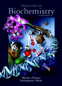
824 Pages • 442,582 Words • PDF • 83.9 MB

743 Pages • 269,880 Words • PDF • 12.4 MB

PDF • 7 MB

45 Pages • 23,141 Words • PDF • 6.2 MB

11 Pages • 9,572 Words • PDF • 623.9 KB

737 Pages • 476,896 Words • PDF • 42.6 MB

393 Pages • 207,369 Words • PDF • 2.5 MB

11 Pages • 10,993 Words • PDF • 2.2 MB

226 Pages • 142,660 Words • PDF • 43.3 MB

449 Pages • 336,317 Words • PDF • 1.9 MB

266 Pages • PDF • 7.5 MB